With the foundation of two women's colleges at the University of Cambridge, in 1869 and 1871, the intellectual life of the city underwent a profound shift. Prior to the 1869, the formal education of Britian's elite had invariably been an all-male affair. The story of women's struggles to be accepted as students and gain recognition within the universities of Oxford and Cambridge has been told many times. Somewhat less attention has however been paid to the ways in which women - despite being excluded from the university proper until the late 1940s - not only participated in, but actually began to influence, the intellectual life of the university from the moment that the colleges were founded. Here, I will concentrate on the Cambridge context to highlight one way in which women influenced elite university life in Britain from the very beginnings of their arrival there. In particular, I investigate how the scientific research of one early college fellow, Marion Greenwood, had rather more influence on physiological research in the university than has hitherto been recognized.
Greenwood arrived at Girton, the first-founded of the two colleges, in 1879. The daughter of a Hull shipping agent, she had attended the Girls’ Grammar School in Bradford before moving to the city. At Cambridge Greenwood found much encouragement for her scientific interests. The Bathurst studentships, founded at Girton's sister college Newnham to support womens’ advanced study in the sciences, were established four years after her arrival, and she found herself the second ever recipient of the award.
Like many women scientists at this time Greenwood had been drawn to microscopy, most likely for its convenience and relative accessibility compared to other equipment-heavy laboratory endeavours. As Marsha Richmond has shown, neither college were able to invest heavily in science at this time, and the women's laboratory that did get founded, the Balfour Laboratory (also by Newnham), was often short of funds. As Greenwood recalled for the Newnham College Letter in 1929, aspiring women physiologists of the 1880s had to be able to adapt to often trying circumstances:
co-education in biological practical work was hardly contemplated, especially in laboratories already over-crowded, and the contrivances by which some amount of practical work was achieved would startle the well equipped student of today. To be the unwelcome tenant of a Bench properly devoted to Chemistry; to carry microscopes thrice weekly into and out of the almost empty room in the Herbarium used for the women's demonstrations in practical Botany; to wrestle with the Pendulum Myograph in a ground floor bathroom of Sidgwick Hall - these experiences have been ours.
Yet despite such hurdles, and an extremely heavy teaching load, Greenwood (who would only halt her physiological research activities after marrying George Parker Bidder in 1899) developed a highly influential line of microscopic research at Cambridge.
 Greenwood’s earliest articles, published in The Journal of Physiology, concerned amoeboid digestion. She described her interest in the digestive processes of amoeba as arising out of the by-then well-known observation that a ‘characteristic’ feature of Rhizopoda was their ‘constant’ ingestion of matter. Whilst many authors had noted the fact that amoeba appeared to ingest material from their surroundings, the processes by which they assimilated it had remained at the margins of microscopic discourse. The vast majority of investigators had, at least since the development of sophisticated fixing techniques during the 1860s, concerned themselves with the processes by which the bodies of cellular organisms might be preserved on slides in order that their anatomy be revealed. In parallel with such studies, chemical analysis of cellular substances – such as those conducted by the German pioneer of biochemistry Justus von Leibig – had begun to detail a conception of microscopic life as both active (giving off materials associated with life in general) and productive (of substances that could be utilized in industrial contexts such as that of the brewing industry). The dramatic results of these had by the 1880s drawn attention away from the classic publications of such figures as Abraham Trembley, Charles Bonnet and Rene Antoine Réaumur, for whom direct observation of living forms had been paramount.
Noting in an article on Rhizopods the existence of ‘but few descriptions which deal at all fully with the mode of ingestion, and fewer which give in detail the changes taking place in ingested bodies’, Greenwood implicitly aligned herself with these eighteenth-century figures. Her commitment to the investigation of cellular activity would lead her to participate in the constitution of a then-emerging conception of amoebae not as inert bodies or substance-producing factories, but as animals species in their own right – as species that could be distinguished by their district activities as well as by their products and fixed anatomical states.
In her Rhizopod studies, Greenwood adopted a technique that in some respects paralleled that developed by the German histologist Robert Koch in 1877. Dissatisfied with what he saw as a lack of reliable depictions of bacteria suspended in their native liquid habitat, Koch had fixed to his microscope slides ‘smears’ of bacteria-containing matter so thin that they presented only a single plane of cellular matter to a microscope lens. From such preparations, Koch had produced photographic depictions of bacterial anatomy that his peers had found deeply convincing. Greenwood would almost certainly have read Koch’s paper, and may well have found in it inspiration for her own studies. Nevertheless, if her techniques were inspired by this source, she adapted them to a very different purpose.
Greenwood created planes of cells not, as did Koch, as prelude to fixing their bodies for observation, but as a means of bringing their living processes into view. Depositing amoeba-carrying water onto a slide, and applying pressure with a coverslip ‘slight enough to allow the emission of short pseudopodia in planes at right angles to the plane of extension’, Greenwood similarly reduced her microscopic subjects to a single surface of analysis. She did not, however, look to stabilize such scenes through fixing and photography. Instead, Greenwood produced with her microscope slides a liquid theatre of cellular activity; a literal two-dimensional stage via which the living drama of the very small might be observed, interacted with, and related.
The animation of Greenwood's theatre of microscopic life required more than simply squeezing organisms into a single plane of existence, however. Greenwood's principal interest in her amoeboid actors did not concern the relationships that they had to one another, but rather the conditions under which they were able to thrive. Central to this concern was consideration of the means by which they assimilated matter from their surroundings. As she noted, though a range of scattered observations existed on the incorporation of outside matter by very simple organisms (most of which took their cue from Christian Gottfried Ehrenberg's famous contentions regarding the presence of 'stomachs' in 'polgastric infusoria'), there had been no systematic study of the processes of amoeboid digestion. In part, this was due to the difficulties involved in identifying the changes that matter underwent once it had been drawn into the bodies of singe-celled animals. By the 1880's, Ehrenberg's contentions regarding the presence of distinct digestive organs in infusoria had been supplanted by the identification of such organisms as unicellular 'protozoa'. Ehrenberg's identification of dark patches in his infusoria as stomachs had attained the status of a cautionary tale amongst histologists. The assimilation by protozoa of nutrients from their surroundings via vacuoles was not accorded great prominence in anatomical discussions more concerned with the extent to which such beings could be identified as possessing clear skin-like borders.
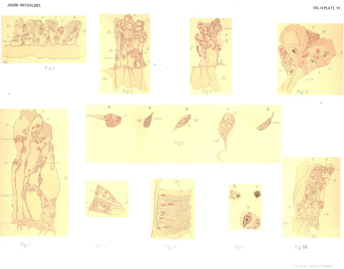
Whereas histological anatomists 'fixed' cells to their slides as a prelude to colouring them with stains, thereby killing them in the process, Greenwood would encourage her objects of study to assimilate coloured matter whilst they were still alive. She portrayed this strategy as having emerged during a chance observation, during which an amoeba formed a vacuole around 'a Monad... and a green Protococcus' (a type of algae, coloured green by chlorophyll). Over a period of six hours, Greenwood had watched the incorporation of the Protococcus into the body of the amoeba, at the end of which the former had retained its green colour. Though it revealed little regarding the difficult-to-observe process of digestion of algae, Greenwood noted, such observations were 'valuable inasmuch as when the digestive vacuole is gone, colour and contour mark them out from the surrounding endosarc [i.e. bodily matter] of the Amoeba, and therefore help to supply some links in the chain of change from ingestion to ejection.' The greenness of the Protococcus meant that its movement through the amoeba could be observed with ease compared to less colourful ingesta. Brightly-tinted material presented her amoeboid actors with props, through the following of which Greenwood might narrate the processes of unicellular digestion.
Over the next decade or so, Greenwood would introduce coloured matter to a wide range of microscopic organisms. In 1888, she applied her dye-feeding technique to a somewhat larger organism: the Hydra. This organism had been studied rather more thoroughly than the amoeba, and so Greenwood found herself negotiating a rather more contentious field of claims relating to its digestive processes. For example, there had been some dispute as to the function of one of the two types of cell found in the body cavity of the Hydra. Though some researchers had suggested these cells functioned as store-houses for nutrients, and others had considered them juvenile forms of the second (and less contentious) type ('vacuolate endoderm' cells). Greenwood supported the conclusions of the latest writers on the topic that the smaller cells were 'glandular' - that is, that they were responsible for the secretion of the Hydra's digestive fluids. By 1894, Greenwood had developed her technique in relation to the infusoria - an article from her research on which would appear in the prestigious Philosophical Transactions of the Royal Society. As Richmond notes, the renowned German physiologist Max Verworn replicated Greenwood's main figure for this piece in the second edition of his Allgemeine Physiologie (1897). He described the study (which he assumed had been conducted by a man) as both ‘very interesting’ and ‘outstanding’. During the late 1880s and early 1890s, Cambridge colleagues and students such as William Bate Hardy, Charles Ballance, and Charles Sherrington cited her studies extensively in their own (subsequently more widely acknowledged) publications.
As already noted, Greenwood was not only a highly original physiological investigator; she also spent a considerable amount of time training a new generation of women scientists at Girton and Newnham. She had initially been somewhat reluctant to take on work that might distract her from her research. On being offered the role of Demonstrator of Physiology at the Balfour laboratory in 1884, she wrote from her parent's house in Oxenhope, Yorkshire to the Mistress of Newnham College Eleanor Sidgwick that:
My first thought would have been about the Studentship and the right [means] of having any time except in the work for which it is given. But that is made unnecessary by the decision of the Committee. Therefore I can only say that while taking the post with some doubt of myself I will do all I can to make the teaching thorough and successful.
I hope it will mean no lessening of research, but only a better arranging of the day's work.
Greenwood nevertheless found that the post required that she invest a considerable amount of energy. In June 1887 she gratefully wrote again to Sidgwick thanking the Balfour Committee for their decision to provide her with assistance in the next academic year. As her collaborator and friend Edith Rebecca (Becky) Saunders would later note:
It is difficult... to appreciate the amount of work involved in teaching... under the conditions which then existed. The courses had to be organized from the beginning. All equipment had to be bought or borrowed, the strictest economy having to be exercised in every way... Had Miss Greenwood not been endowed with a splendid constitution she could hardly have accomplished all that was required and have carried out research at the same time.
Whether or not Greenwood's interest in the digestive processes of minute organisms contributed to her constitutional strength must remain nat present a matter of speculation. It is certainly the case, however, that her studies had a profound (if all-too-little-acknowledged) influence on physiological science during the 1890s.

- No links match your filters. Clear Filters
-


 CitesC.A. Ballance and C.S. Sherrington, 'On Formation of Scar-Tissue', Journal of Physiology 10 (6), (1889), pp. 550-578.
Description:'Cambridge colleagues and students such as William Bate Hardy, Charles Ballance, and Charles Sherrington cited her [Greenwood's] studies extensively in their own more widely acknowledged publications.'
-
 CitesM. Greenwood to E. Sidgwick, 3rd June 1887.
Description:'the post [of Demonstrator of Physiology at the Balfour Laboratory] required that she [Greenwood] invest a considerable amount of energy. In June 1887 she gratefully wrote again to Sidgwick thanking the Committee for their decision to provide her with assistance in the next academic year.'
-
CitesM. Greenwood, 'On Digestion in Hydra, with some Observations on the Structure of the Endoderm', Journal of Physiology 9 (5-6) (1888), pp. 317-344.
Description:'Greenwood would introduce coloured matter to a wide range of microscopic organisms. In 1888, she applied her dye-feeding technique to a somewhat larger organism: the Hydra. This organism had been studied rather more thoroughly than the amoeba, and so Greenwood found herself negotiating a rather more contentious filed regarding its digestive processes. For example, there had been some dispute as to the function of whether one of the two types of cell found in the body cavity of the Hydra. Though some researchers had suggested these cells functioned as store-houses for nutrients, and others had considered them juvenile forms of the second (and less contentious) type ('vacuolate endoderm' cells), Greenwood supported the conclusions of the latest writers on the topic that these cells were 'glandular' - that is, that they were responsible for the secretion of digestive fluids.'
-
CitesM. Greenwood, 'On the Constitution and Mode of Formation of "Food Vacuoles" in Infusoria, as Illustrated by the History of the Processes of Digestion in Carchesium polypinum', Philosophical Transactions B 185 (1894), pp. 355-383.
Description:'By 1894, Greenwood had developed her [dye-feeding] technique in relation to the infusoria - an article from her research on which would appear in the prestigious Philosophical Transactions of the Royal Society. The renowned German physiologist Max Verworn replicated Greenwood's main figure for this piece in the second edition of his Allgemeine Physiologie (1897) noting that the study (which he assumed had been conducted by a man) was both ‘very interesting’ and ‘outstanding’.'
-
CitesM. Verworn, Allgemeine Physiologie: ein Grundriss der Lehre vom Leben (2nd ed.) (Jena, 1897).
Description:'By 1894, Greenwood had developed her technique in relation to the infusoria - an article from her research on which would appear in the prestigious Philosophical Transactions of the Royal Society. The renowned German physiologist Max Verworn replicated Greenwood's main figure for this piece in the second edition of his Allgemeine Physiologie (1897) noting that the study (which he assumed had been conducted by a man) was both ‘very interesting’ and ‘outstanding’. '
-
CitesM.L. Richmond, '"A Lab of One's Own": The Balfour Laboratory for Women at Cambridge University', Isis 88 (3) (1997), pp. 422-455.
Description:'As Marsha Richmond has shown, neither college were able to invest heavily in science at this time, and the women's laboratory that did get founded, the Balfour Laboratory (also by Newnham), was often short of funds.'
'As Richmond notes, the renowned German physiologist Max Verworn replicated Greenwood's main figure for this piece in the second edition of his Allgemeine Physiologie (1897).'
-
CitesW.B. Hardy and L.B. Keng, 'On the Changes in the Number and Character of the Wandering Cells of the Frog induced by the presence of Urari or of Bacillus Anthracis', Journal of Physiology 15 (4) (1893), pp. 361-400.
Description:'Cambridge colleagues and students such as William Bate Hardy, Charles Ballance, and Charles Sherrington cited her [Greenwood's] studies extensively in their own more widely acknowledged publications.'
-
QuotesE.R. Saunders, 'Mrs G.P. Bidder (Marion Greenwood)', Letter [Newnham College Roll] (January 1933).
Description:'the post [of Demosntrator of Physiology at the Balfour Laboratory] required that she [Greenwood] invest a considerable amount of energy... As her collaborator and friend Edith Rebecca (Becky) Saunders would later note:
It is difficult... to appreciate the amount of work involved in teaching... under the conditions which then existed. The courses had to be organized from the beginning. All equipment had to be bought or borrowed, the strictest economy having to be exercised in every way... Had Miss Greenwood not been endowed with a splendid constitution she could hardly have accomplished all that was required and have carried out research at the same time.
-
QuotesM. Greenwood Bidder, 'History of the Balfour Laboratory', Letter [Newnham College Roll] (January, 1929), pp. 45-47.
Description:'co-education in biological practical work was hardly contemplated, especially in laboratories already over-crowded, and the contrivances by which some amount of practical work was achieved would startle the well equipped student of today. To be the unwelcome tenant of a Bench properly devoted to Chemistry; to carry microscopes thrice weekly into and out of the almost empty room in the Herbarium used for the women's demonstrations in practical Botany; to wrestle with the Pendulum Myograph in a ground floor bathroom of Sidgwick Hall - these experiences have been ours.' (45)
-
QuotesM. Greenwood to E. Sidgwick, 2 July 1884.
Description:'Greenwood was not only a highly original physiological investigator; she also spent a considerable amount of time training a new generation of women scientists at Girton and Newnham. She was initially been somewhat reluctant to take on work that might distract her from her research. On being offered the role of Demonstrator of Physiology at the Balfour laboratory in 1884, she wrote to the Mistress of Newnham College Eleanor Sidgwick that:
My first thought would have been about the Studentship and the right [means] of having any time except in the work for which it is given. But that is made unnecessary by the decision of the Committee [of the Balfour Laboratory]. Therefore I can only say that while taking the post with some doubt of myself I will do all I can to make the teaching thorough and successful.
I hope it will mean no lessening of research, but only a better arranging of the day's work.
She nevertheless found that the post required her to invest a considerable amount of energy.'
-
QuotesM. Greenwood, 'On the Digestive Process in some Rhizopods', Journal of Physiology 7 (3) (1886), pp. 253-273.
Description:'Greenwood’s earliest articles, published in The Journal of Physiology, concerned amoeboid digestion. She described her interest in the digestive processes of amoeba as arising out of the by-then well-known observation that a ‘characteristic’ feature of Rhizopoda was their ‘constant’ ingestion of matter. Whilst many authors had noted the fact that amoeba appeared to ingest material from their surroundings, the processes by which they assimilated it had remained at the margins of histological discourse.'
'Noting in an article on the Rhizopods the existence of ‘but few descriptions which deal at all fully with the mode of ingestion, and fewer which give in detail the changes taking place in ingested bodies’, Greenwood implicitly aligned herself with the founding figures of histology. Her commitment to the investigation of cellular activity would lead her to participate in the constitution of a then-emerging conception of amoebae not as inert bodies or substance-producing factories, but as animals species in their own right – as species that could be distinguished by their district activities as well as by their fixed anatomical states.'
'Greenwood's principal interest in her amoeboid actors in these did not concern the relationships that they had to one another, but rather the conditions under which they were able to thrive. Central to this concern was consideration of the means by which they assimilated matter from their surroundings. As she noted, though a range of scattered observations existed on the incorporation of outside matter by very simple organisms (most of which took their cue from Christian Gottfried Ehrenberg's famous contentions regarding the presence of 'stomachs' in 'polgastric infusoria'), there had been no systematic study of the processes of amoeboid digestion.'
'Whereas histological anatomists 'fixed' cells to their slides as a prelude to colouring them with stains, thereby killing them in the process, Greenwood would encourage her objects of study to assimilate coloured matter whilst they were still alive. She portrayed this strategy as having emerged during a chance observation, during which an amoeba formed a vacuole around 'a Monad... and a green Protococcus' (a type of algae, coloured green by chlorophyll). Over a period of six hours, Greenwood had watched the incorporation of the Protococcus into the body of the amoeba, at the end of which the former had retained its green colour. Though it revealed little regarding the difficult-to-observe process of digestion of algae, Greenwood noted, such observations were 'valuable inasmuch as when the digestive vacuole is gone, colour and contour mark them out from the surrounding endosarc [i.e. bodily matter] of the Amoeba, and therefore help to supply some links in the chain of change from ingestion to ejection.' The greenness of the Protococcus meant that its movement through the amoeba could be observed with ease compared to less colourful ingesta. Brightly-tinted material presented her amoeboid actors with props, through the following of which Greenwood might narrate the processes of unicellular digestion.'
-
QuotesM. Greenwood, 'On the Digestive Process in some Rhizopods. Part II.', Journal of Physiology 8 (5) (1887), pp. 263-310.
Description:'In her Rhizopod studies, Greenwood adopted a technique that in some respects paralleled that described one developed by the German histologist Robert Koch in 1877. Dissatisfied with what he saw as a lack of reliable depictions of bacteria suspended in their native liquid habitat, Koch had fixed to his microscope slides ‘smears’ of bacteria-containing matter so thin that they presented only a single plane of cellular matter to a microscope lens. From such preparations, Koch had produced photographic depictions of bacterial anatomy that his peers had found deeply convincing. Greenwood would almost certainly have read Koch’s paper, and may well have found in it inspiration for her own studies. Nevertheless, if her techniques were inspired by this source, she adapted them to a very different purpose.'
Greenwood created planes of cells not, as did Koch, as prelude to fixing their bodies for observation, but as a means of bringing their living processes into view. Depositing amoeba-carrying water onto a slide, and applying pressure with a coverslip ‘slight enough to allow the emission of short pseudopodia in planes at right angles to the plane of extension’, Greenwood similarly reduced her microscopic subjects to a single surface of analysis. She did not, however, look to stabilize such scenes through fixing and photography. Instead, Greenwood produced with her microscope slides a liquid theatre of cellular activity; a literal two-dimensional stage via which the living drama of the very small might be observed, interacted with, and related.'
-
-
-