- External URL
- Creation
-
Creators (Definite): William Bate Hardy; Lim Boon KengDate: 1893
- Current Holder(s)
-
- No links match your filters. Clear Filters
-
Cites
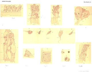
 Plate XI, Journal of Physiology 15 (4) (1893). Figs. 1-12 from W.B. Hardy and L.B. Keng, 'On the Changes in the Number and Character of the Wandering Cells of the Frog induced by the presence of Urari or of Bacillus Anthracis'.
Description:Explanation of Plate XI (figs. 1-12):
'Fig. 1. Amphophile condition of the granules of an eosinophile cell. Nucleus swollen and showing no structure. Third day after injection of urari, methylene blue. Oc. 4, ob. 1/12th cam. luc.
Fig. 2. Binuclear eosinophile cell, stained with methylene blue. Third day after urari. Oc. 4, ob. 1/12th cam. luc.
Fig. 3. (a) Dead and disintegrating eosinophile cell. (b) Dead and disintegrating hyaline cell, methylene blue. Third day after urari. Oc. 4, ob. 1/12th cam. luc.
Fig. 4. Binuclear hyaline cells, stained with methyl green acidulated with acetic acid. Urari 20 hours. Oc. 4, ob. E, Zeiss, cam. luc.
Fig. 5. Hyaline cell laden with amorphous ingestion, methylene blue. (a) 20 hours after urari. Oc. 4, ob. Ith cam. luc. (b) 24 hours after injection of anthrax. Oc. 4, ob. F, Zeiss, cam. luc.
Fig. 6. Hyaline cell with vacuolated protoplasm. Active ingestion going on, methylene blue, 24 hours after anthrax. Oc. 4, ob. 1/12th cam. luc.
Fig. 7. Hyaline cell which has ingested two eosinophile cells and whose protoplasm is studded with eosinophile granules. Methylene blue 24 hours after injection of anthrax. Oc. 4, ob. 1/12th cam. luc.
Fig. 8. Hyaline cell which bas ingested an eosinophile cell. Film preparation stained with eosin and Loeffler's methylene blue, after urari. Oc. 4, ob. 1/12th cam. luc.
Fig. 9. Hyaline cell with the remains of another cell occupying a vacuole. Methylene blue. Oc. 4, ob. 1/12th cam. luc.
Fig. 10. Cell from peritoneal cavity of a Frog 16 hours after injection into that cavity of methylene blue dissolved in normal salt solution. Drawn when alive.
Fig. 11. Cell from the lymph of the foot of the same animal.
Fig. 12. Cells from the lymph of the foot of the same animal four hours after a second injection of methylene blue into the peritoneal cavity. Living cell drawn.' (373-374)
Fig. 1 in text:
'Stage I. Changes in the eosinophile cells. The first effect of the poison is to modify the granulation of the eosinophile cell so that it stains with basic dyes. In other words, an amphophile granulation is produced. The granules at the same time increase slightly in size and become spherical and less refractive (Fig. 1). The granules of the normal cell are usually slightly spindle-shaped and very lustrous. The cells at the seat of inoculation (always the dorsal lymph sac in our experiments) show this change more completely than those obtained elsewhere but, if the dose be sufficiently large, amphophile cells will be found in the lymph from other parts of the body. In many of these amphophile cells, especially in those which retain this modified granulation the longest, the nucleus loses its lobed character and swells up to form a structureless sphere.' (365)
'In any one cell the spheres are of different sizes and often present varying stages in the digestive act, and they are composed of material which stains deeply with a solution of methylene blue, so that a fully loaded cell in such stained preparations may bear some resemblance to an eosinophile cell in the amphophile condition, especially to those in which the lobed nucleus has swollen and to form a sphere. The similarity however is only superficial, for the balls of ingested material are of different sizes and each lies in a definite digestive vacuole hollowed out of the cell, whereas the amphophile granules are quite or nearly of the same size and are evenly distributed throughout the cell substance. The nuclei too present very striking differences; that of the amiphophile cell may be of the typical lobed form, or it may be swollen and spherical. In the second case we are probably dealing with a dying cell, for the nucleus shows no trace of structure but stains uniformly with methylene blue and presents a curious glassy appearance (Fig. 1).' (367-368)
Figs. 7 and 8 in text:
'The dissolution of cells spoken of above which marks the leucocytosis produced by the presence of microbic poisons or of urari is effected in two ways. The cells may simply break up in the plasma,... At a later period, in what we shall speak of as the second stage, the dissolution is no longer of this passive kind but certain of the cells are actively destroyed by the hyaline cells. The eosinophile cells in particular are destroyed by being bodily ingested by the hyaline cells, and this fate overtakes them while they are apparently quite normal so far as their nuclei and granules are concerned. In this way there come to be introduced into the body of a great number of the hyaline cells large masses of eosinophile granules which, as digestion of the prey proceeds, are scattered as isolated granules through the cell substance of the hyaline cells (Figs. 7 and 8). Thus at this stage cells are found which possess the same nuclear characters and have the same type of cell body and of movement as the hyaline cells but which also show an eosinophile granulation, and in film preparations suich cells appear, so far as their structural features are concerned, intermediate in character between the normal eosinophile and the normal hyaline cell.' (362-363)
Fig. 7 in text:
'The ingestion of the eosinophile cells is carried out with considerable vigour, so that one finds not infrequently a hyaline cell which contains an eosinophile cell still intact and obviously only lately ingested occupying one vacuole, another eosinophile cell in a more advanced stage of digestion in a second vacuole, and remains of other cells, notably the granules, occupying vacuoles scattered through the rest of the cell substance. One such stained with methylene blue is shown in Fig. 7, and a cell with an intact eosinophile cell in a vacuole is shown at Fig. 8 as seen in a film preparation.' (372)
'The later stages of the leucocytosis following the introduction of anthrax into the Frog. The earlier phenomena manifested by the wandering cells of the frog when anthrax is present have been described elsewhere'. In the communication referred to they were followed up to the point where the bacilli are destroyed and the lymph is left with a large number of eosinophile, hyaline and basophile (rose reacting) cells. Both the eosinophile and hyaline cells at this period are actively amoeboid. The further phenomena resemble those described above as constituting the resolution of the leucocytosis produced by the presence of urari, that is to say active ingestion and destruction of the eosinophile cells by the hyaline cells occurs. An instance of this, obtained from the lymph of a Frog 24 hours after inoculation with anthrax, is shown at Fig. 7.' (373)
Figs. 10-12 in text:
'if methylene blue dissolved in normal salt solution be injected into a Frog (e.g. into the peritoneal cavity), the lymph in all parts of the body will be found to contain cells more or less laden with what appears to be solid blue spheres. Figures 10 and 11 represent such cells as they appear when alive. If at this period a second injection be made, then four hours afterwards the cells will present the appearance shown in Fig. 12. In other words, the vacuoles have been reconstituted round the already absorbed pigment and the fluid they contain is very obviously tinged with the dye.
In the case of the cells represented at Fig. 12 both injections were made into the peritoneal cavity and the lymph examined was obtained from the foot.' (370)
-
Cited by
 An Amoeboid Theatre: Marion Greenwood Bidder's physiological research at Cambridge (1879-1899)
Description:'Cambridge colleagues and students such as William Bate Hardy, Charles Ballance, and Charles Sherrington cited her [Greenwood's] studies extensively in their own more widely acknowledged publications.'
-
Cited by
 M. Greenwood and E.R. Saunders, 'On the Rôle of Acid in Protozoan Digestion', Journal of Physiology 16 (5-6) (1894), pp. 441-467.
Description:'We believe that the presence or absence of a blue colour in the vacuole depends on the predominance of one or the other of two antagonistic processes, the first setting free methylene blue as the proteid which it impregnates is dissolved, the second depositing the pigment so freed round the residual solid nidus of the vacuole. A comparable deposition of methylene blue from solution has been described in the wandering cells of the frog [note: 'W. B. Hardy and Lim Boon Keng. This Journal, XV. p. 370, 1893.']; it must not be forgotten, however, that methylene blue is not readily soluble in solutions of salts and that there is reason to believe that salts are present in a digestive vacuole. The absence of colour which is certainly striking in Carchesium may, then, have complex causes.' (458-459)
-
Quoted by
 T. Quick, Minute Mediation: Cell Physiology, Print-Making and Industry in Late Victorian Cambridge
Description:'The concerns of two authors centred on the identification and classification of wandering cells in frogs. As with Hardy’s paper on crustacean skin, however, it was not so much the classification of cell-types that the article focused on, but the extent to which different cell-types could be identified as arising out of a single ancestral progenitor – could be, in fact, different manifestations of the same fundamental cell-type. Central to this investigation, as with Balance and Sherrington’s publication, was the possibility that one cell-type might possess the capacity to morph into another. Though wandering cells in frogs are ‘not only sharply marked off from one another by their respective structural characteristics, but also in the way that they behave when foreign substances… are present in the plasma’, there could nevertheless, Lim and Hardy suggested, ‘be no finality in the view’ that each type enjoyed ‘complete independence’.[1] Strikingly, and in sharp contrast with Hardy’s single-authored paper, their conclusions asserted differences between types of cell, and especially their lack of interchangeability: ‘no real loss of identity was observed; nothing was seen which suggests the formation of an eosinophile from a hyaline cell or vice versa’ Indeed, the two had observed ‘the devouring of eosinophile cells by the hyaline cells… One cannot conceive on the hypothesis that the eosinophile and hyaline cells represent merely phases in the development of the same cell, why the one should devour the other’.[2]
To reach their conclusions, Lim and Hardy had looked to the same materials that had animated Greenwood’s studies. But rather than persuade their Crustacean subjects to ingest particles of dye, Lim and Hardy injected it into the bloodstream of the animals directly.'


