- External URL
- Creation
-
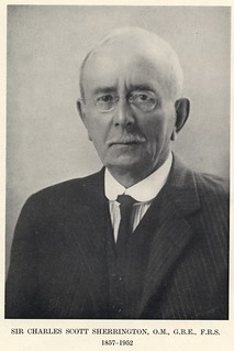
Creators (Definite): Sir Charles Alfred Ballance; Sir Charles Scott SherringtonDate: Oct 1889
- Current Holder(s)
-
Holder (Definite): The Journal of Physiology
Concerns the repetition of the ‘classic’ experiments in tissue formation by Zeigler. Includes a series of photographs taken using a glass Ziegler chamber for the study of tissue formation - describes equipment used, method employed (esp. re: requirements for specimens amenable to observation at higher powers of magnification).
Animals used: rabbits and guinea-pigs.
Preparations either examined fresh, or after being subjected to hardening and staining processes. Hardened using osmic acid (either 5% water solution or vapour from a 1% solution) - includes description of method used. Preparations mounted in ‘Farrant’s gum solution or, after dehydration, in xylol balsam’. Sometimes after-stained with ‘picrocarmine, with fuchsin, methylene blue, eosin, or most frequently with haematoxylin (Ehrlich's solution).’
Notes that the creation of ‘black particles’ is due to use of ‘shellac glue’ in the preparation process (p. 560).
Describes a second chamber method: ‘Two chambers (or four) were placed side by side in the subcutaneous tissue. At the end of twenty-two hours they were taken out, one was dropped into osmic acid, and the other was sealed with warm paraffin.’ (p. 565) - also describes the use of acid, the method used for ensuring isolation of chamber,
Contributors not named as authors: ‘we were... much assisted in the interpretation of the appearances of the osmic fixed preparations by the processes described by Miss M. Greenwood for the Rhizopoda.’ (‘This Journal’, Vol. vii. p. 253, Vol. viii. p. 263.)
Illustrations:
Fig 1: Rabbit - contents of chamber after 72hrs in peritoneal cavityt. Prepared over osmic acid vapour.
Fig. 2: Rabbit - content of chamber after 18hrs. Prepared over osmic vapour.
Fig. 3: Guinea-pig - content of chamber after 3 days in subcutaneous tissue. Osmic acid solution, ‘Erlich’s logwood’.
Fig. 4: Rabbit - same chamber as (1).
Fig. 5: Same as (1).
Fig. 6: Rabbit - content of chamber after 72hrs in peritoneal cavity. Osmic acid vapour.
Fig. 7: As fig (6)
Fig 8: Guinea-pig - content of chamber after 8 days in subcutaneous tissue. Osmic acid vapour.

- No links match your filters. Clear Filters
-


-
-

-

-
CitesM. Greenwood, 'On the Digestive Process in some Rhizopods', Journal of Physiology 7 (3) (1886), pp. 253-273.
Description:'We doubt whether without very special apparatus the cells of the tissues of mammalia can be kept in sufficiently normal condition for sufficient length of time to compass observations on ingestion by living cells; we were however much assisted in the interpretation of the appearances of the osmic fixed preparations by the processes described by Miss M. Greenwood for the Rhizopoda. Her observations [note: 'This Journal, Vol. VII. p. 253, Vol. VIII. p. 263.'] were conducted on living specimens of Amoeba proteus and Actinosphaerium, and she was able to follow in these animals under the microscope all the visible phenomena accompanying the ingestion of prey. In our preparations we had as it were a number of amoebae, many of which had been actively engaged in ingesting living prey, immediately before the reagent had been used that killed them so rapidly as to allow no time for any great departure from their previous aspect.' (559)
'leucocytes served... as a pabulum for the active plasma-cells [note: 'The plasma-cells are considered by Metschnikoff... ' (note cut short here)]. Just as, in the extremely interesting observations given by M. Greenwood [note: 'This Journal, Vol. VII. p. 253. Vol. VIII. p. 263.'], little monads, Euglenae and Algae coexisting in the same water with Amoeba proteus were by it ingested, so leucocytes become the prey of the plasma-cell, and are by it included and ingested. And if the growth and proliferation of the plasma-cells be of importance in the process of repair, what circumstance more propitious than the presence in abundance of nutriment so delicately adapted and so highly organized as the substance of the leucocytic cell? Of Amoeba and Actinosphaerium it was remarked that the food most suitable to these forms is unshielded non-coagulated proteid matter. A low degree of vitality, a diminished activity of its protoplasm, renders an organism easier prey, more readily captured and more readily absorbed. The plasma-cell may in some respects be taken as a hothouse variety of amoeba; it finds its unshielded non-coagulated proteid in the dead or dying leucocyte.' (568-569)
-
CitesM. Greenwood, 'On the Digestive Process in some Rhizopods. Part II.', Journal of Physiology 8 (5) (1887), pp. 263-310.
Description:'We doubt whether without very special apparatus the cells of the tissues of mammalia can be kept in sufficiently normal condition for sufficient length of time to compass observations on ingestion by living cells; we were however much assisted in the interpretation of the appearances of the osmic fixed preparations by the processes described by Miss M. Greenwood for the Rhizopoda. Her observations [note: 'This Journal, Vol. VII. p. 253, Vol. VIII. p. 263.'] were conducted on living specimens of Amoeba proteus and Actinosphaerium, and she was able to follow in these animals under the microscope all the visible phenomena accompanying the ingestion of prey. In our preparations we had as it were a number of amoebae, many of which had been actively engaged in ingesting living prey, immediately before the reagent had been used that killed them so rapidly as to allow no time for any great departure from their previous aspect.' (559)
'leucocytes served... as a pabulum for the active plasma-cells [note: 'The plasma-cells are considered by Metschnikoff... ' (note cut short here)]. Just as, in the extremely interesting observations given by M. Greenwood [note: 'This Journal, Vol. VII. p. 253. Vol. VIII. p. 263.'], little monads, Euglenae and Algae coexisting in the same water with Amoeba proteus were by it ingested, so leucocytes become the prey of the plasma-cell, and are by it included and ingested. And if the growth and proliferation of the plasma-cells be of importance in the process of repair, what circumstance more propitious than the presence in abundance of nutriment so delicately adapted and so highly organized as the substance of the leucocytic cell? Of Amoeba and Actinosphaerium it was remarked that the food most suitable to these forms is unshielded non-coagulated proteid matter. A low degree of vitality, a diminished activity of its protoplasm, renders an organism easier prey, more readily captured and more readily absorbed. The plasma-cell may in some respects be taken as a hothouse variety of amoeba; it finds its unshielded non-coagulated proteid in the dead or dying leucocyte.' (568-569)
-
 CitesPlate XXXI, Journal of Physiology 10 (6) (1889). Figs. 1-5 from C.A. Ballance and C.S. Sherrington, 'On Formation of Scar-Tissue'.
Tags: osmic acid, logwood, osmic vapour
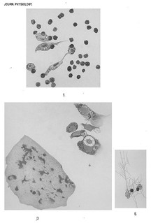
Description:Explanation of Plate XXXI (figs. 1-5):
'Fig. 1. Contents of experimental chamber that had remained 72 hours in the peritoneal cavity of the rabbit. Five large amoeboid plasma-cells, with altered red corpuscles and apparently dead leucocytes. Outlined with camera lucida. Apochromatic oil immersion and ocular No. 4. Zeiss. Prepared over osmic vapour.
Fig. 2. Contents of a chamber for 18 hours in the peritoneal cavity (rabbit); near the centre of the chamber. Fibrin filaments, leucocytes, red corpuscles, and an ill-defined granular mass forming a nodal point in the fibrinous network-the beginning of a "cell-islet." Outlined under camera. Similar method of preparation, and similar magnification to preceding.
Fig. 3. Fragment of inflammatory membrane formed within a chamber placed for three days in the subcutaneous tissue (guinea-pig). Islets and groups of islets scattered through the membrane. Zeiss, Obj. A, Oc. 2. Osmic acid solution, and Ehrlich's logwood.
Fig. 4. Contents of same chamber as in Fig 1. Close to the opening of the chamber. Five plasma-cells, one of them continuing a leucocyte within a large vacuole. Magnification and method of preparation as in Fig. 1.
Fig. 5. Contents of same chamber. Two plasma-cells and two red corptiscles; the plasma-cells are indistinguishably united with fine filaments of fibrin in their surrounding, some of which are given in the figure. Osmic acid vapour. Zeiss, apochr. system, oc. No. 2.' (575)
Figs. 1 and 4-5 in text:
'The preparations gave an almxiost bewildering number of examples of the infinite variation in shape of the large amoeboid plasma-cells, which also varied very considerably in size, and as to granules. The body of the cell was for the most part plate-like, being in many instances extended into so thin a film that its exact limit was hard to determine, especially when, as occasionally happened, the granules of the cell-body were less pronounced towards the periphery. Some idea of the wide diversity of outline exhibited by individual cells may be gathered from our figures. Cf Figs. 1, 4, 5, 6, 7 and 8, Plates XXXI. and XXXII.' (558)
'In the specimens obtained from chambers that had rested for seventy-two hours in the subcutaneous tissue of the guinea-pig, we found individuals among the plasma-cells, which showed wellmarked vacuolation, Figs. 1, 4, 5, Pl. XXXI. For the most part the matter within the vacuole was a granular debris that furnished no sufficient clue as to its nature. But in a few it was indisputable that the vacuole contained, more or less altered but still perfectly easily recognisable, a leucocyte or red blood corpuscle. In Fig. 4 is shown the appearance presented by one of these cells. A large vacuole contains a somewhat faintly stained body, which is finely granular and indistinctly nucleated. It is a little smaller than is the nucleus of the plasma-cell itself. Fine threads seemed to pass from the sides of the vacuole across the cavity to the substance of the included leucocyte. Taken with the context afforded by examination of other cells in the neighbourhood we believe that this and other similar instances were examples of leucocytes lying in vacuoles in the plasma-cells. Many stages of ingestion could be found. Cf. Figs. 1, 4, 5, Pl. XXXI. Simple approximation, the hollowing out of a little bay in the side of the plasma-cell into which the leucocyte was as it were drawn, partial inclusion, total inclusion - all these were exemplified.' (559)
Figs. 1 and 5 in text:
'Contiguous plasma-cells or even those a little distance apart were often connected together by their processes (Figs. 1, 5, 7 and 8, Plates XXXI. and XXXII.). The bands of connection might be short thick arms or long gossamer threads of protoplasm. By similar arms and threads the cells seemed to adhere to the most diverse objects in their surrounding. The surface of the cover-glass, a filamllent of fibrin, a hair, a fibre of cotton, a lump of the cement fastening the sides of the chamber together, all afforded points to which the processes from the plasma-cells would cling (Figs. 14 and 15).' (560)
Fig. 1 in text:
'Here must be mentioned another sign of degeneration in the leucocytes examined in these chambers [note: 'Kuss, Paris, 1846. Paget, Surgical Pathol. p. 151.']. Many of them showed the triple and multiple nuclear bodies that are universally regarded as evidence of the lethal disintegration of the nucleus - as Fleming names it, the "fragmentation" of the nucleus. On the other hand the cell-body of the leucocyte was not granular or fatty, but fairly evenly though deeply tinted by the osmium. These points are seen in Fig. 1, Plate XXXI.' (556-557)
Figs. 2-3 in text:
'Eighteen Hours.- In chambers removed after the appearance of fibrin within them, but before the stay within the body had exceeded eight and forty hours, it was usual to find a number of areas in which leucocytes were present in much greater numbers than elsewhere. Fig. 2, Pl. XXXI.
The tendency to collect to certain points which the leucocytes evinced in even very early specimens was more marked in these later preparations. About the nodal points of the fibrinous network crowds of them were present. The ouitlying individuals were frequently arranged in lines along the converging filaments of fibrin. The older within certain limits these films of coagulum the more obvious the aggregation of the leucocytes into certain groups. For convenience on account of their prominence. and apparent importance in subsequent stages we have been accustomed to refer to these groups shortly as the cell-islets. Cf. Fig. 3, Pl. XXXI. They are little collections of cells, occurring constantly, scattered about in the thin cellular membranes which grow over and within the glass chambers.' (557)
-
 CitesPlate XXXII, Journal of Physiology 10 (6) (1889). Figs. 6-13 from C.A. Ballance and C.S. Sherrington, 'On Formation of Scar-Tissue'.
Tags: osmic acid, logwood
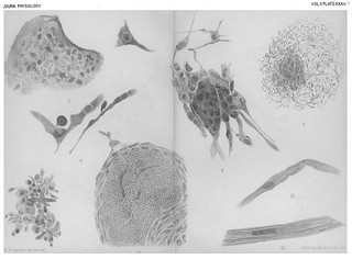
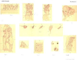
Description:Explanation of Plate XXXII (figs. 6-13):
'Fig. 6. Giant cells from chamber 72 hours in the peritoneal cavity (rabbit). Zeiss apochr. system, ocul. 5. Osmic acid vapour.
Fig. 7. Plasma-cells from same preparation which furnished Fig. 6.
Fig. 8. " Cell islet " from inflammatory film obtained in a chamber left eight days in the subcutaneous tissue of the guinea-pig, At the margin it is united to outlying plasma-cells. Zeiss oil, oc. 4. Osmic acid vapour.
Fig. 9. Young cicatricial tissue of anastomosing branched cells, some of which are represented under the higher magnification in Fig. 17. From a thrombosed artery (syphilis) near the centre of the thrombus. Zeiss A, oc. 3. Logwood. Preparation kindly shown us by Dr Seymour Sharkey.
Fig. 10. "Cell islet" from inflammatory membrane obtained from chamber five days in the peritoneal cavity of the rabbit. Osmic acid solution. Zeiss oil imm. and oc. 2.
Fig. 11. Mass of blood-cells (? clot) surrounded by fibroblastic cells, and invaded by them at four places. Inflammatory membrane from chamber eight days in subcutaneous tissue. Magnification as in preceding, and prepared in similar manner.
Fig. 12. Fusiform plasma-cell (fibroblast) surrounded by a fibrillated material which forms a thread-like band of connective tissue. Zeiss oil and oc. 4. Osmic vapour. From chamber 10 days in subcutaneous tissue.
Fig. 13. Similar but larger and thicker fibrous band from same preparation. Similar preparation and magnification.' (575-576)
Figs. 6-8 in text:
'The preparations gave an almxiost bewildering number of examples of the infinite variation in shape of the large amoeboid plasma-cells, which also varied very considerably in size, and as to granules. The body of the cell was for the most part plate-like, being in many instances extended into so thin a film that its exact limit was hard to determine, especially when, as occasionally happened, the granules of the cell-body were less pronounced towards the periphery. Some idea of the wide diversity of outline exhibited by individual cells may be gathered from our figures. Cf Figs. 1, 4, 5, 6, 7 and 8, Plates XXXI. and XXXII.' (558)
Fig. 6 in text:
'There were present also in chambers of eighteen hours', twentytwo hours', twenty-six hours', forty-eight hours', and seventy-two hours' standing, as also in others of older date containing well formed granulation tissue, many giant cells (Fig. 6) - huge multi-nucleate cells, that obviously in many instances were cell-fusions. ' (560)
Figs. 7-8 in text:
'Contiguous plasma-cells or even those a little distance apart were often connected together by their processes (Figs. 1, 5, 7 and 8, Plates XXXI. and XXXII.). The bands of connection might be short thick arms or long gossamer threads of protoplasm. By similar arms and threads the cells seemed to adhere to the most diverse objects in their surrounding. The surface of the cover-glass, a filamllent of fibrin, a hair, a fibre of cotton, a lump of the cement fastening the sides of the chamber together, all afforded points to which the processes from the plasma-cells would cling (Figs. 14 and 15).' (560)
Figs. 8-9 in text:
'In membranes of ten, fourteen, and even eighteen days' growth, not all the cells nor even the majority were spindle-shaped. A vast number were triradiate, and multiradiate; some had but one process; very few were rounded. Many recalled to mind the branched fixed corpuscles of the cornea. Long tapering branches united cell to cell, not only the cells of one plane one with another, but the cells of different planes also (Figs. 8, 9 and 17). A meshwork of infinite variety and complexity was thus established. But in all these examples of plasma cells in the stable as well as in the previously described labile forms, the granular nature of the cell substance and the clear oval nucleus were characters never lost.' (563)
Figs. 8 and 10 in text:
'in the specimens of more than forty-eight hours' duration, the plasma-cells begin to apply themselves to the islet-groups of leucocytes. Cf. Figs. 8 and 10. They surround the leucocytes. The islets come to consist of a central portion made up of leucocytes, and an outer zone of large and granular plasma-cells. In this way the islets seem to increase rapidly in size. Neighbouring islets appear to become merged together.' (561)
Fig. 11 in text:
'It was among the plasma-cells of the fringe of the islets that we noticed the earliest regularly fusiform cells, the immediate precursors of fibrous elements in the new tissue. It is true that plasma-cells of an irregular spindle-shape were observable not rarely among even the earliest of the plasma-cell swarm entering the chamber. But in those instances the outline was probably but one of many which the amoeboid cell successively assumed, and generally it was not of the same character as the regularly fusiform type prevailing among these plasma-cells in the outskirts of an islet. In that latter the majority of the cells lay in lines concentrically set about a core of ill-stained, broken-down matter that composed the centre of the mass. Cf. Fig. 11, Pl. XXXII. The fusiform fibroblasts began in fact the encapsulation of the débris of the breaking-down blood cells, &c.' (526)
Figs. 12-13 in text:
'Older specimens revealed further progress in the formation of a fibrous-tissue membrane. After a stay of eight days, or ten days, or fourteen days in the subcutaneous tissue in many instances the islets consisted of plasma-cells alone. The leucocytes had disappeared. The pigmented remnants of the red blood corpuscles were much longer traceable. In many places along certain lines the spindle-shaped cells had become attenuated, and formed distinct bands and often long and delicate cords (Figs. 12, 13). In many places in the tenth day specimens, and in some of the eighth day ones an inter-cellular substance showing fibrillation exists (Fig. 12).' (562)
-
 CitesPlate XXXIII, Journal of Physiology 10 (6) (1889). Figs. 14-19 from C.A. Ballance and C.S. Sherrington, 'On Formation of Scar-Tissue'.
Tags: haemotoxylin, osmic acid
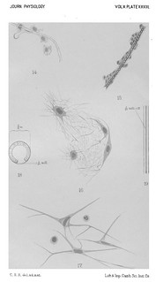
Description:Explanation of Plate XXXIII (figs. 14-19):
'Fig. 14. From chamber five days in subcutaneouis tissue. Plasma-cells adhering to a cotton-fibre. Osmic vapour and carmine. Zeiss apochlr. oil and oc. 4.
Fig. 15. From chamber eight days in subcutaneous tissue. Plasma-cells adhering to a hair, which had accidentally been allowed to get into the wound. Zeiss obj. D, oc. 2. Osmic acid solution and haematoxylin.
Fig. 16. Inflammatory membrane from chamber eight days in the abdominal cavity; taken from a tenuous portion of the membrane. Four fibroblasts, in a film which is composed of an extremely irregularly arranged network of filaments resembling fine fibrin threads. The processes from the cell-body are continuous apparently with the fibrils of the matrix. Osmic acid vapour and haematoxylin. Zeiss apochr. oil imm. and oc. 4. Outlined with camera lucida.
Fig. 17. Stellate fibroblasts and two leucocytes from same preparation as Fig. 9, more highly magnified. Zeiss apochr. oil and oc. 4. Outlined with camera lucida.
Fig. 18. The modified Ziegler chamber; the sketch shows the actual size employed.
Fig. 19. Portion of the chamber seen edgewise, showing the opening between the cover-glasses. Enlarged 12 times.' (576)
Figs. 14-15 n text:
'Contiguous plasma-cells or even those a little distance apart were often connected together by their processes (Figs. 1, 5, 7 and 8, Plates XXXI. and XXXII.). The bands of connection might be short thick arms or long gossamer threads of protoplasm. By similar arms and threads the cells seemed to adhere to the most diverse objects in their surrounding. The surface of the cover-glass, a filamllent of fibrin, a hair, a fibre of cotton, a lump of the cement fastening the sides of the chamber together, all afforded points to which the processes from the plasma-cells would cling (Figs. 14 and 15).' (560)
Fig. 16 in text:
'Each individual cell was of a discoid or fusiform figure, and granular, with a large clear nucleus. The edge of the disc was thin and often deeply scalloped; it merged, under all methods of staining used by us, at certain points quite imperceptibly, in a tenuous film which composed the bulk of the membrane proper. When fixed with osmic acid and after-stained with haematoxylin (Ehrlich's), this membrane is shown to contain, if not to be entirely made up of, a feltwork of filaments, like filaments of fibrin. These cross in every direction in the plane of the membrane, without prominent arrangement in any one particular sense. The individual filaments vary a good deal in size. Fig. 16, P1. XXXIII.' (561-562)
Fig. 18 in text:
'Two circular cover-glasses, each 5/8 of an in. in diameter and .006 of an in. in thickness, were fastened together so as to form a little flat glass chamber, in the manner employed by Ziegler. A strip of tinfoil placed between them at their edge along 11/12 of their circumference was cemented by shellac on each face to the corresponding surface of the cover-glass. The tiny chamber thus formed had therefore between the two ends of the strip of tin-foil an opening into the interior. The tin-foil first employed was 1/10 mm. thick; that thickness was inconvenient, as the depth of the chamber was then too great for higher powers of the microscope to explore. Tin-foil 1/20 mm. in thickness was subsequently employed. With this thickness membranes were obtained between the cover-glasses that made very satisfactory microscopical specimens. Fig. 18, Plate XXXIII.' (552-553)
-
 CitesSir Seymour John Sharkey
Description:From explanation of Plate XXXII:
'Fig. 9. Young cicatricial tissue of anastomosing branched cells, some of which are represented under the higher magnification in Fig. 17. From a thrombosed artery (syphilis) near the centre of the thrombus. Zeiss A, oc. 3. Logwood. Preparation kindly shown us by Dr Seymour Sharkey.' (575)
-
 Cited by
 An Amoeboid Theatre: Marion Greenwood Bidder's physiological research at Cambridge (1879-1899)
Description:'Cambridge colleagues and students such as William Bate Hardy, Charles Ballance, and Charles Sherrington cited her [Greenwood's] studies extensively in their own more widely acknowledged publications.'
-

-
Quoted byT. Quick, Minute Mediation: Cell Physiology, Print-Making and Industry in Late Victorian Cambridge
Description:Ballance and Sherringon cited Greenwood’s work extensively in their 1889 paper.
we were… much assisted in the interpretation of the appearances of the osmic fixed preparations by the processes described by Miss M. Greenwood for the Rhizopoda… In our preparations we had as it were a number of amoebae, many of which had been actively engaged in ingesting living prey, immediately before the reagent had been used that killed them so rapidly as to allow no time for any great departure from their previous aspect.[1]
And again:
Just as, in the extremely interesting observations given by M. Greenwood, little monads, Euglena and Algae coexisting in the same water with Amoeba proteus were by it ingested, so leucocytes become the prey of the plasma-cell, and are by it included and ingested.[2]
Sherrington and Ballance’s success on the prominent stage of one of the first truly international medical congresses was no doubt due in considerable degree to their own skill and dedication to their work. Yet Ballance’s dramatic recollection of his and Sherrington's ‘conversion’ of Zeigler to their conception of leukocyte function obscures the broader context from which their study arose. Greenwood’s work not only informed the topic of study of the pair, but provided them with a conceptual lens through which they interpreted their wandering cells. Ballance and Sherrington's performance in Germany had at least in part been written, if not directed, by their Girton and Newnham colleague.
-
-
-


