- External URL
- Creation
-
Creator (Definite): Marion Greenwood BidderDate: 1892
- Current Holder(s)
-
- No links match your filters. Clear Filters
-
Cites
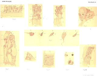
 Plate IX, Journal of Physiology 13 (3-4) (1892). Figs. 1-10 from M. Greenwood, 'On Retractile Cilia in the Intestine of Lumbricus Terrestris'.
Tags: picrocarmine, osmic vapour, corrosive sublimate
Description:Explanation of Plate IX (figs. 1-10):
'In all the figures
g.c. = gland cell.
i.c. = ciliated cell.
a.c. = wall of alimentary canal.
typ. = typhlosohle.
y.c. = yellow cells of ecelomic epithelium (chloragoge Zellen).
m.f. = muscular fibres.
nuc. = nucleus.
gran. = secretory granules.
f.g. = ingested particles of fat.
ex. = masses of substance, possibly excretory in nature, found especially in long hunger.
Fig. 1. Transverse section through the gut of Lumbricus at a point where the typhlosohle is complex. The figure is quite diagrammatic and is merely intended to show the general relations of the lining epithelium and the presence and distribution of yellow cells.
Fig. 2. (In this figure and in the following figures, unless special statements are made, the magnification was that effected by Zeiss, Oc. 3, Obj. F.)
Gland cell holding secretory granules and showing basal homogeneous substance (hardened).
Fig. 3. Gland cell, secretory granules dissolved.
Fig. 4 a, b. Taken from different regions of the walls of the alimentary canal of one earthworm. To show variation in the height of the cilia.
c. To show long cilia passino out from block-like end of cells. (Hardened in Flemming's fluid.)
d. To show vacuole in ? badly-noturished ciliated cell, and division of cell substance into internal processes.
e. Ciliated cell after long hunger. (d and e macerated in 30 p.c. alcohol.)
Fig. 5. From typhlosohle, fasting worm. To show marked ciliation and occurrence of secretory granules in gland cells. (Corrosive sublimate preparation.)' (258-259)
Fig. 1 in text:
'The circular muscle fibres which succeed are again arranged in bundles, and the connective tissue which separates them and which surrounds the abundant neighbouring blood vessels is continuous with that which forms a support for the innermnost layer - the intestinal epithelium. In the cavity of the gut, as I have said above, definite glandular recesses are absent, but through the greater part of its length a well marked involution of the dorsal wall into the intestinal cavity forms the typhlosohle (Pl. IX., Fig. 1. typ.). In this ridge all the layers I have just named are represented; thus it is a highly vascular structure, and yellow cells like those which form the external coat of the gut almost fill its concavity.' (241)
'This intestinal epithelium is, I believe, only one layer in depth. Claparède [note: ' Claparede, Zeit. fur wiss. Zool. Bd. XXXIX'], it is true, speaks of many layers, but his figure is too diagrammatic to allow one to believe that he had seen everything in a section that later methods show. Vejdovsky [note: 'Fr. Vejdovsky, op. cit.'] too, in a figure of Lumbricus, represents small cells as lying at the base of the columnar cells which make up the adult epithelium and evidently regards them as "ersatzzellen." I have not however been able to establish their occurrence at all constantly in Lumbricus. Nuclei indeed are seen besides those which, belonging to the ciliated cells, are placed half-way down the mucous membrane and beside the basal line of nuclei which may be recognized in the gland cells (Fig. 1), but they seem irregular in numnber and occurrence, and I am inclined rather to regard them as belonging to the subepithelial connective tissue or to rare in-wandering leucocytes.' (248)
Figs. 2-3 in text:
'The secretory cells noticed and figured by Vejdovsky [note: 'Fr. Vejdovsky, loc. cit.'] and Benham [note: 'Benham, loc. cit.'] recall the unicellular glands which have already been described in Hydra [note: 'C. Jickeli, Morphol. Jahrb. Bd. VIIL; M. Greenwood, This Journal, Vol. IX.']; they taper slightly towards the internal surface of the gut and more strikingly towards their points of attachment (Figs. 2, 3). These cells display conspicuously, at least at times, a nucleus, cell suibstance and secretory granules; the granules are so numerous in the typically fasting state that it is difficult to realize then the existence of other cell constituents. The protoplasm, when under these conditions it is made evident, is seen to stretch as a supporting framework or spongework throughout the cell; at such times as the granules are less numerous, a basal part of the cell substance, holding the nucleus, is free from them (Fig. 2). When accumulated thus or when it stretches as a temporary network through the cell, the protoplasm shows, I believe, no further obvious structural differentiation. The secretory granules are preserved by osmic vapour, and admirably by corrosive sublimate; they are however more easily broken down by reagents than are the remarkably resistant mucous granules of the unicellular glanids of the skin.' (242-243)
Figs. 4-5 and 7 in text:
''It may be urged, by those who accept the records of these observers, that the minute variations I have described are not incompatible with the received conception of a ciliated cell, or are even artificial and due to want of uniformity in preparation. I must say then that after constant treatment the appearances vary, and that in two other points there is yet a more mainifest deviation from type. In the first place, a vacuole is sometimes present in the external part of the protoplasm (i.e. under the basal band) (Fig. 4 d), while the same region may be collapsed or folded after inaceration so that the cell has a constricted neck. The vacuole is often wanting, however, and the cell substance under the action of hardening reagents, as distinguished from those that macerate, is cominonly dense and homogeneous (Figs. 5, 7, i.c.).' (245-246)
Figs. 4-5 in text:
'there is a comparatively long stretch of intestinal wall in the posterior half of the animal's body where the majority of the cells are in active ciliary movement in the fresh state and where in the hardened specimen the cilia spring from the blocked end of a cell or from a typical basal band (Fig. 4 a, b, c). These are present alone as far sometimes as the level of the ending of the typhlosohle, at other times at this point scattered gland cells are striking. I may perhaps repeat that here and always when glands are present the extreme outer edges of the ciliated cells are apparently in contact laterally in the fresh state, and when hardened show at best only inconspicuous openings between them. Thus the secretion of the cells which lie behind has to reach the cavity of the intestine by narrow channels recalling those by which the mucous glands of the skin open outwards [note: 'J. N. Langley, loc. cit.'] (Fig. 5).' (249)
Fig. 4 in text:
'Cells with obvious cilia. (1) Certain cells, save that they are unusually elongated and branch internally, are not far removed in structure from such ciliated cells as characterize the buccal mucous membrane of Triton, that is their cilia, uniform in length, spring from an apparently hyaline band.
(2) But in many cases the height of the cell is disproportionately increased (Fig. 4 d.), the filiform processes into which its substance breaks internally are very striking and the basal band takes the form of a triangular plug of hyaline substance, the base of the triangle being the apparent point of origin of the cilia while the apex passes off gradually into the protoplasm of the cell (Fig. 4 c.).
(3) And yet again cilia may be traced through the external zone and are only lost when they pass into the protoplasm beyond. With this arrangement the basal band seems sometimes to be made up of rods stouter than the cilia, which look very like their intracellular prolongations; at other times however it is hyaline save for the striae which are the necessary expression of the existence of the perforating cilia. The cilia occasionally bear tiny varicosities before they pass into the body of the cell (Fig. 4 d). Under a sufficiently high power these are distinguishable as belonging each to a ciliary thread [note: 'These varicosities stand singly as a rule, but I think it is possible that each cilium at times bears two, one at the outer limit and one at the inner limit of the basal band.'], and they recall Heidenhain's description of similar thickenings which may be seen under suitable conditions at the base of the intestinal rods of the dog.' (245)
'It will be gathered from the indirect nature of the evidence which I have brought forward that the most convincing proof of the existence of retractile cilia is wanting; I have not seen a hyaline border put out protoplasmic processes, nor have I seers any complete retraction of cilia after death. Variations in length may occur in any one earthworm (Fig. 4 a, b), and in the median region of the typhlosohle, where I think this special form of mobility in the cells is developed characteristically, the cilia, when they show, are finer, more scattered, and less vigorous in action than on the gut walls.' (255)
Figs. 5 and in text:
'It is, I think, indisputable that the typhlosohle is ciliated at times (Fig. 5); the edges of certain cells lining the alimentary canal may, on the other hand, be smooth; now since the typhlosohle has a hyaline border in all well-nourished worms belief in retractility seems inevitable unless we suppose that there is even extraordinary individual variation in structure. It is very probable that when Metschnikoff [note: ' E. Metschnikoff, Zool. Anzeiger, 1878. 1. Jahrgang.'] speaks of the ciliated intestinal epithelium of Microstomum lineare as illustrating a type of cell which has quite lost the power of taking up solid nourishment, he formulates a view which is held widely, yet here in Lumbricus we have cells now ciliated, now crowded with fat. I would urge that these cells are not typical, but rather transitional between the absorptive cells of Hydra with their retractile flagella and the epithelium with its border of rods which is figured in the frog and in the mammal. They are extensile and mobile to a degree which is even surprising when we remember the firm union of many vertebrate tissues, and apparently definite displacement of their substance may occur. Thus there is at different times attenuation or expansion of the outer part of the body of each cell or a vacuole may be present; while not rarely the lateral outlines are so muich obscured that the cells seem confluent in transverse section (Fig. 9). But it is in the border that there is special power of change; the cell would seem indeed to have at its disposal a certain amount of substance which inay form a hyaline band giving origin to fine cilia, or may on the other hand be arranged in rods, stouter, but less readily discernible. And when the cell is especially narrow this changing mass of substance deepens from without inwards; it is wedge-shaped rather than zone-like. '. (235)
Figs. 6-8 in text:
'Cells without obvious cilia. The forms which I place in this group have no motile protoplasmic processes projecting freely into the intestine. In the fresh state the external edges of adjacent cells touch and give the effect of a continuous band, rarely a double band, of hyaline substance (Fig. 8) from which little bullke may be separated off by pressure. In hardened specimens the homogeneity is sometimes preserved (Fig. 7), but as a rule it is possible to apply treatment which brings out certain structural complications. Thus when macerated cells are mounted in fluids of low refractive power it is usual to see that the greater part of the free border is made up of rods set side by side, only a narrow internal zone being still hyaline (Fig. 6 d). The rods remind one irresistibly of Heidenhain's [note: 'R. Heidenhain, op. cit. Taf. 1, figs. V. VI. VII. VIII.'] description of the "Stübehenorgan" in the vertebrate intestine: they are not always equally obvious, nor is their share in the formation of the whole border of the cell constant. Thus at times the internal hyaline band takes up half the depth of the wvhole border; at other times the distinction into two layers is not clear (Fig. 6 b) and the rods are so little separated that the cell seems to have a coarsely striated edge. The inner homogeneous band stains deeply with picro-carmine: the rods stain almost as fairntly as do actuial cilia.' (246-247)
Fig. 6 in text:
'in the great majority of specimens fat is present in large amount and this at once mnarks out the cells which hold it. The fat drops vary much in size, slightly in shape ; the smallest globules lie immediately beneath the free edge of the cell; larger drops are crowded more internally so that protoplasm and nucleus may be obscured, and, apparently passing down, they certainly distend the filiform internial processes (Fig 6 a, b, c).' (247)
'The fat globules which may crowd the typhlosohlar epithelium in some regions, and may be present, though less markedly, in the walls of the alimentary canal, are invariably most abundant in animals taken from rich earth or from leaf mould (Fig. 6 a, b, c). During periods of hunger they disappear, tarrying internally to the nucleuis and apparently passing down the filiform internal processes of the cell which they distend.' (252)
Fig. 10 in text:
'Up to this point, in writing of the gut of Lumbricus I have dealt with epithelium cells and their contents, but there are generally collections of substances between the cells which are both striking and puzzling. These have the form of complex oval masses of matter, and the nature of their constituents is at once variable and obscure. Among these constituents are amorphous clumps of proteid matter (Fig. 10 c.), and irregular particles coloured yellow or orange in the fresh state and staining black with osmic acid, while in some cases there is a pigmented substance which resists the action of caustic potash. I have never demonstrated the presence of nuclei, and indeed if these masses are to be looked upon as cellular, the cells of which they are made up are much changed and disorganized. Fluid is present in varying amount (cp. Fig. 10 d with Fig. 10 b), and the actual shape of any mass is determined by the surrounding epithelium cells, or by the viscosity of some of its proper constituents.' (256)


