- External URL
- Creation
-
Creator (Definite): Sir Charles Scott SherringtonDate: 1893
- Current Holder(s)
-
- No links match your filters. Clear Filters
-
Cites
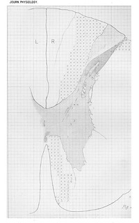
 Plate XIII, Journal of Physiology 14 (4-5) (1893). Figs. 1-4 from C.S. Sherrington, 'Note on the Spinal portion of some Ascending Degenerations'.
Description:Fig. 1 in text:
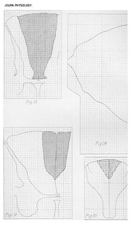
'Microscopical Examination. At the level of the lowest point of the scar there is (Pl. XIII., Fig. 1) a wedgeshaped area of degeneration with sclerosis at the outer free angle of the lateral division of the posterior column, involving nearly the whole width of the division of the column, and extending into the depth of the column as far as the apex of the substantia gelatinosa of the posterior horn. The sclerosis and the degeneration one millimeter higher is most dense in the portion abutting on the fissure between the lateral and posterior columns, and in a small part of that region no sound fibres at all are to be found.' (256)
Fig. 2 in text:
'One millimeter higher. The width of the area of degeneration and sclerosis has at the base of the lateral-posterior column become greater, but it still does not pass into the median-posterior column. The area extends through the entire depth of the lateral-posterior column. The part adjoining the fissure between the lateral and posterior column contains no healthy nerve-fibres. The scar is here attached for a slight extent to the subpial membrane of the cord in front of the fissure, and the most posterior edge of the cerebellar tract, where it lies against the entrance of the posterior root, is degenerated; a few healthy fibres existing among the degenerated. There is no obvious sclerosis here. One millimeter higher (Fig. 2). The width of the area of degeneration and sclerosis has at the base of the lateral-posterior column become greater, and reaches the outer edge of the median-posterior column. It passes through the entire depth of the lateral-posterior column. In the region abutting on the septum between the lateral and posterior column no healthy nerve-fibres are seen.' (256-257)
Fig. 3 in text:
'Just below the pyramidal decussation (Fig. 3). The degeneration in the most lateral part of the lateral posterior column is very severe; degeneration in that column occupies the whole width of its more superficial part, but becomes of slight intensity next to the septum dividing the column from the median posterior. The area of the whole right hand posterior column being taken as 415 (weight in lead foil), the portion of it containing signs of degeneration amounts to 267, and of this portion in an area of 57 the degeneration is very severe, approaching absolute.' (257)
Fig. 4 in text:
'At the level of the upper part of the pyramlidal decussation (Fig. 4) where the decussation of the fillet exists well marked, the area of degeneration in the right funiculus cuneatus in shape and extent differs little from that at the level just described. It has however a broader line of contact with the underlying grey matter at the base of what was the cornu posterius.' (258)
-
Cites
 Plate XIV, Journal of Physiology 14 (4-5) (1893). Figs. 5-6 and 11-13 from C.S. Sherrington, 'Note on the Spinal portion of some Ascending Degenerations'.
Description:Fig. 5 in text:'Level close above the pyramidal decussation; lower end of the nucleus hypoglossi. (Fig. 5.) The main degreneration, that in the funic. cuneatus, still presents the same general features as before. Although occupying nearly as wide a portion of the surface, it has become separated from] the middle line by three times as great a distance as before. It extends much more deeply into the grey matter here than lower down. At this level grey mnatter pervades the deeper portion of the funiculus in scattered masses.' (258)
Fig. 6 in text:
'Immediately below the point at which the central canal opens into the floor of the lVth ventricle and where the central grey matter has already come well to the surface the degeneration has the following extent. (Fig. 6.)' (259)
Fig. 11 in text:
'The distribution of that portion of the white matter still completely sound, in which at least no degeneration has been detected by myself after search in a large series of specimens is a matter of some interest. Its details can be gathered from Fig. 11. Part of it is that portion of the white matter of the deep part of the lateral column which lies between the dorsal edge of the lateral horn of grey matter and the ventral limb of the substantia gelatinosa of the posterior horn, and a small area abutting laterally on the above. Part of it is the portion of the lateral column abutting on the lateral edge of the grey matter of the anterior horn; part of it the portion of the anterior column abutting on the mesial edge of the anterior horn.' (273-274)
Fig. 12 in text:
'In the middle of the field of scattered degeneration there is however in each posterior column a considerable islet of very severe or almost absolute degeneration. The topographical relations are seen in analysis Fig. 12. If the area of the posterior columns be 636 then of this 411 is occupied by absolute degeneration. In 11 squares no signs of degeneration are detectable, all these eleven squares abut on the grey cornua.' (275)
-
Cites
 Plate XV, Journal of Physiology 14 (4-5) (1893). Figs. 7-10 from C.S. Sherrington, 'Note on the Spinal portion of some Ascending Degenerations'.
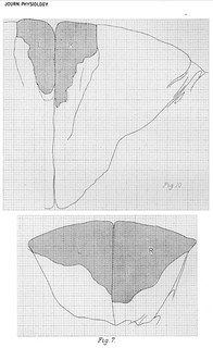
Description:Fig. 7 in text:
'The right degeneration therefore diminished in size more rapidly between the 10th thoracic and the 5th thoracic roots than did the left degeneration. The part then of the right hand degeneration at 10th thoracic level (Fig. 7) which does not correspond with the left must consist of fibres that do not in so great proportion as do fibres of the rest of it contribute to the formation of Goll's column in the cervical region.' (271)
Fig. 10 in text:
'Microscopical examination. Seat of lesion. A small mass of fibrous tissue continuoous with the dura mater entered as it were the-posterior column and penetrated into it for quite two thirds of its depth. It occupied the entire width of the column passing from the bundles of the entering left hand posterior root to those of the right hand root; it certainly implicated the root bundles in part at least. The portion of the posterior column not inivolved was an area the shape and extent of which is seen in Fig. 10.' (263-264)
'After ascending to near the level of the 3rd cervical nerve the size of the area of degeneration (Fig. 10) is found to have again undergone considerable reduction, especially compared with the large posterior column of these high levels of the cord. It has become diminished in width, so that to it now belongs less than half as much of the free edge of the posterior column as at the level of the 6th thoracic.' (270)
-
Cites
 Plate XVI, Journal of Physiology 14 (4-5) (1893). Figs. 14-17a from C.S. Sherrington, 'Note on the Spinal portion of some Ascending Degenerations'.
Description:Fig. 11 in text:
'An area of almost absolute degeneration skirts the posterior limb of the substantia gelatinosa; between this deep zone of degeneration and the great superficial area there is a zone of less severe degeneration, in which numbers of healthy nerve-fibres exist, both coarse and fine of various degrees of size; very few of the fibres are quite large, none of them so large as the fibres of the cerebellar tract; the majority are fine, averaging less than 2 µ. The area of absolute degeneration contains a scanty number of nerve-fibres still in process of degeneration, but the bulk of it is composed of cicatricial tissue in which numerous spaces represent the places previously occupied by nerve-fibres. The topography of the degeneration here can best be gathered from fig. 11.' (272)
Figs. 15-16 in text:
'The posterior column which is much larger in area than at the last level examined contains some obvious scattered degeneration in its deepest part, that nearest to the grey matter. About 60-70 degenerating fibres can be found thLere in each section (Fig. 15). The area of absolute degeneration is less wide from right to left than it is lower down the cord. Its area is not really much less than at the level of the 8th cervical (231-254), but relatively to the whole posterior column instead of being a fifth part of it it is little more than a tenth.
At the level of the 2nd cervical nerve-root (Fig. 16) the posterior column of the cord although much larger than at the 2nd thoracic is not so large as at the 5th cervical. Yet the area of absolute degeneration in it is not only smaller absolutely than at the last mentioned level, but it is relatively smaller also, i.e. considerably less than one-tenth of the total column instead of somewhat more than one-tenth. Its edge is at least as sharp as previously. It is somewhat compressed laterally, so that it occupies less than a sixth of the free edge of the column. It is conterminous for the greater part of its side with a small septum. The area of the posterior column outside the degenerated area does not contain an obvious amount of scattered degeneration. There are indeed eight fibres in it that appear to be degenerated, but more I cannot detect; the position of these fibres is marked on the analysis map.' (280-281)
Fig. 16 in text:
'It is instructive to compare the configuration of the ascending degenerations at this level with the descending degeneration examined at the same level in the same species four months after a very large cortical lesion involving more than the whole "cord-area" of the cortex outside the gyrus fornicatus (Fig. 16 A). In the Macacque the crossed pyramidal tract extends outside its main mass into a peripheral zone described by France [note: 'Philos. Transacts. vol. B. 1889.'] and by myself [note: 'This Journal, March, 1889.']; in fcetus of the Macacque I find this peripheral sheet acquire myelin at the same epoch as does the pyramidal tract elsewhere; between this peripheral sheet (which in some specimens reaches halfway down the thoracic region) and the main body of the tract is an interval occupied almost exclusively by a coarse-fibred (megalomitous) tract, this is shown to degenerate (Fig. 16) upwards, and is the direct cerebellar. Although there is considerable overlapping of the territories of the ascending and descending degenerations the converse character of the general picture offered by the two degenerations is unmistakeable. The distinction between the ventral part of the lateral reticular formation and the part occupied by 'pyramid' fibres is sharp; in the foetus by paucity of myelin in the latter, in the adult by the ventral containing small fibres (micromitous), the latter fibres of very various sizes (poikilomitous).' (281-282)
Fig. 17 in text:
'The anatomy of the degeneration existing below the site of the lesion deserves some mention. The transverse lesion was at the 10th thoracic segment and involved the whole extent of the cord. A preparation taken below the level of origin of the 11th thoracic root about 17 mm. below the lesion is well comparable with a preparation from a level between the 9th and 8th roots because the anatomy of the normal cord is in the two places only slightly dissimilar. The antero-lateral column has a heavy degeneration throughout the greater portion of its dorsal half. The distribution of this is shown by the deeper shading in Fig. 17.' (282)
'A comparison is interesting between this descending degeneration two segments below a transverse spinal lesion and the descending degeneration of the pyramidal tract at the same level resulting from ablation of the whole "cord region" of the cortex (except Schäfer and France's gyrus fornicatus area). Not only is the area of descending degeneration (Fig. 19) in the lateral column much more extended in the case of the spinal lesion than in that of the cortical (Fig. 17 A) but within the area itself the density of the degeneration is much greater. Commingled therefore with cerebial fibres descending from the "cord-region" of the cortex are, as well as ascending spinal fibres, fibres which descend and are probably of spinal, some possibly of cerebellar (Marchi) [note: 'Rivista Sperim. di Freniatria, 1888.'] origin, all of them contained within the field appropriated by nomenclature to the crossed pyramidal tract [note: 'Sherrington. This Journal, 1885.'].' (282)
'There is another area of heavy degeneration in the most ventral portion of the anterior column, and along the margin of the ventral median fissure. A scattered degeneration of much slighter intensity involves the whole of the rest of the antero-lateral column, except for a narrow zone immediately abutting upon the grey substance. This narrow zone is narrowest at the free apex of the lateral horn and at the extreme ventral tip of the anterior horn; it is widest in the inlet of white matter bounded ventrally by the lateral horn dorsally by the base of the posterior horn, and it is fairly wide at the bottom of the anterior column. In each posterior colunmi a small oval patch of degeneration lies imbedded deeply, the mesial end nearly meeting the border of the grey matter, its other end directed toward the place of entrance of the posterior root (Fig. 17).' (283)
-
Cites
 Plate XVII, Journal of Physiology 14 (4-5) (1893). Figs. 18-22 and 24 from C.S. Sherrington, 'Note on the Spinal portion of some Ascending Degenerations'.
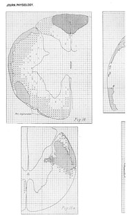
Description:Figs. 18-22 in text:
'The diminution in the quantity of degeneration is well marked in Rhesus as already shown. In man in a cord the opportunity of preparing sections from which I owe to Dr Hadden, after complete destruction of the posterior columns near the level of the 6th thoracic root the ascending degeneration in the posterior column showed a similar continued diminution in passing along the cervical region. (Figs. 18, 19).
[table here enumerates proportions of degenerated columns]
In the Dog in result of complete transverse lesion at the level, of 9th thoracic root the ascending degeneration after 45 days gave the following measurements. (Figs. 20, 21.)
[table here enumerates proportions of degenerated columns]
The decrease in the area of degeneration is therefore marked in the dog, but apparently not so great as in Rhesus.' (284)
Fig. 19 in text:
'A comparison is interesting between this descending degeneration two segments below a transverse spinal lesion and the descending degeneration of the pyramidal tract at the same level resulting from ablation of the whole "cord region" of the cortex (except Schäfer and France's gyrus fornicatus area). Not only is the area of descending degeneration (Fig. 19) in the lateral column much more extended in the case of the spinal lesion than in that of the cortical (Fig. 17 A) but within the area itself the density of the degeneration is much greater. Commingled therefore with cerebial fibres descending from the "cord-region" of the cortex are, as well as ascending spinal fibres, fibres which descend and are probably of spinal, some possibly of cerebellar (Marchi) [note: 'Rivista Sperim. di Freniatria, 1888.'] origin, all of them contained within the field appropriated by nomenclature to the crossed pyramidal tract [note: 'Sherrington. This Journal, 1885.'].' (282)
Fig. 22 in text:
'In the experiment from which the above measurements were quoted the degeneration at the 6th thoracic root level measured between a quarter and a third of the whole column, instead of more than a half as in Rhesus. In an experiment in which the cord (Dog) was completely divided at the level of the 6th thoracic the condition of the cord three segments higher is seen in analysis in Fig. 22. By measurement it was found that
[table here enumerates proportions of degenerated columns]
i.e. less than a quarter instead of more than a half as in Expt. III. The degeneration had been allowed 52 days.' (285)
Fig. 24 in text:
'Weigert-Pal preparations. At the level of the 2nd thoracic root 49 squares were counted in the median posterior column of the right side and 323 in the column of the left side. The 49 squares contained 2317 nerve-fibres, the 323 squares 14891 nerve-fibres; the 49 right hand squares gave an average therefore of 47.2 nerve-fibres per square, the 323 an average of 46.1 per square. (Fig. 23.)
At the level of the 2nd cervical root 49 squares in approximately the same part of the column as the 49 taken at the 2nd thoracic root contained 2394 nerve-fibres, i.e. 488 fibres per square. In the other column, i.e. right hand, 196 squares were counted in a position more closely abutting upon the dorsal angle of the column and more closely hugging the median dorsal septum than the squares counted at the 2nd thoracic level, in order to allow for the kind of shifting of position to which the fibres in the column are subjected as revealed by the degenerations studied above. (Fig. 24.) In the 196 squares 9724 fibres were found, giving an average of 49.6 to the square. The condensation found therefore would account for a diminution in size of the area of absolute degeneration of about 8 p.c.' (288)
-
Cites
 Plate XVIII, Journal of Physiology 14 (4-5) (1893). Figs. 23 and 25-31 from C.S. Sherrington, 'Note on the Spinal portion of some Ascending Degenerations'.
Description:Fig. 23 in text:
'Weigert-Pal preparations. At the level of the 2nd thoracic root 49 squares were counted in the median posterior column of the right side and 323 in the column of the left side. The 49 squares contained 2317 nerve-fibres, the 323 squares 14891 nerve-fibres; the 49 right hand squares gave an average therefore of 47.2 nerve-fibres per square, the 323 an average of 46.1 per square. (Fig. 23.)
At the level of the 2nd cervical root 49 squares in approximately the same part of the column as the 49 taken at the 2nd thoracic root contained 2394 nerve-fibres, i.e. 488 fibres per square. In the other column, i.e. right hand, 196 squares were counted in a position more closely abutting upon the dorsal angle of the column and more closely hugging the median dorsal septum than the squares counted at the 2nd thoracic level, in order to allow for the kind of shifting of position to which the fibres in the column are subjected as revealed by the degenerations studied above. (Fig. 24.) In the 196 squares 9724 fibres were found, giving an average of 49.6 to the square. The condensation found therefore would account for a diminution in size of the area of absolute degeneration of about 8 p.c.' (288)
Figs. 25-26 in text:
'An obvious objection to this interpretation of the occurrence of "geminal fibres " is that where numerous degenerate fibres are present it is only in accordance with probability that in some cases two will happen to lie alongside each other. Against the appearance being thus explicable as a mere chance juxtaposition, it has to be remembered that the fibres referred to are not merely juxtaposed, but are also as judged of by the tint that they assume under various staining methods, and by the degree of formal disintegration which they exhibit, in the same stage of decay one as another (Fig. 25); and further that in some instances they exhibit the possession of a sheath common to both of the two fibres (Fig. 26).' (295)
Fig. 26 in text:
'Although the fibres of the pyramidal tract are in the cord largely commingled with other fibres, in the pyramid itself its fibres are not commingled with others except with arciform, easily distinguishable by their course in a plane at right angles to that of the pyramidal fibres. At the same time not quite all the fibres of one pyramid degenerate after even very large ablations from the cortex of one hemisphere. I attempted therefore to obtain a very large and complete degeneration of the pyramid and to then see if the few sound fibres remaining in the degeneration mass occurred in pairs; in fact one attempted to obtain a positive picture of the fibres in place of the negative already furnished. Search made in this way reveals both in Rhesus (Fig. 26) and in Dog numerous examples of "geminal" arrangement among the sparse sound fibres in a severely degenerated pyramid, in fact most of the larger of the sound fibres occur in pairs, a few in triplets.' (295-296)
Fig. 30 in text:
' it might be expected that "geminal" fibres just as they degenerate together and escape degeneration together, might together myelinate. This is in fact the case. Fibres myelinating in pairs are numerous in the developing cord. Fig. 30 represents an instance from the human cord. The pairs both small and large are numerous in the developing cord, and especially so the larger.' (297)
-
Cited by
 C.S. Sherrington, 'On the Anatomical Constitution of Nerves of Skeletal Muscles; with Remarks on Recurrent Fibres in the Ventral Spinal Nerve-root', Journal of Physiology 17 (3-4) (1894), pp. 210-258.
Description:'The actual number of root-ganglion nerve-fibres in an ordinary muscular nerve-trunk is considerable. I have in not a few instances counted them, using the same method of counting as for the fibres of the spinal cord [note: 'This Journal, XIV. p. 287.' (224-225)
Sherrington's 1893 paper also highlights the anatomy of the posterior spinal root considered in detail in 'On the Anatomical Constitution':
'the above observations contribute an item toward illustration of an arrangement indicated by many researches that bear upon the afferent entries of the cord, namely, that a posterior spinal root establishes direct connections ('adjunctions' to judge by the results of the Golgi method) on its own side of the median plane not with one but with a great number of the chain of segments of the neural axis (e.g. afferent fibres from the leg plunging directly into grey matter of brachial spinal segments), whereas evidence of such direct relation of the fibres of a motor root to more than one or some very limited number of spinal segments is still wanting or at present even negatived.' (300)
-
Cited by
 Charles S. Sherrington and 'Mechanical Objectivity'.
Description:'the mode in which Sherrington presented his [early] work owed as much to the formulae, tables and calculations of the Cavendish physicists as it did to the anatomic accuracy of his Cambridge mentors. Rather than present lavish illustrations of his nerve investigations, as had Gaskell (fig. 1), Sherrington supplied tables of response measurements, graphs of reaction-intensities, and introduced grids with which he might reduce the complexity of nervous anatomy (fig. 2).'
-
Cited by
 T. Quick, 'Disciplining Physiological Psychology: Cinematographs as Epistemic Devices, 1897-1922', Science in Context 30 (4), pp. 423-474.
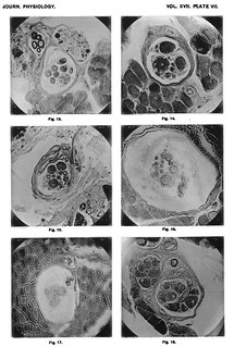
Description:'the mode in which Sherrington presented his work owed as much to the formulae, tables and calculations of the Cavendish physicists as it did to the anatomic accuracy of his Cambridge mentors. Rather than present lavish illustrations of his nerve investigations, as had Gaskell, Sherrington supplied tables of response measurements, graphs of reaction-intensities, and introduced grids with which he might reduce the complexity of nervous anatomy (fig. 1). His study addressing the pioneering histological work of Angelo Ruffini replaced the latter's all-encompassing hand-drawn delineations with a series of microphotographs (fig. 2) (Sherrington 1893; Sherrington 1894).'

