- Creation
-
Creators (Definite): Sir Charles Scott Sherrington; The Cambridge Engraving CompanyDate: 1893
- Current Holder(s)
-
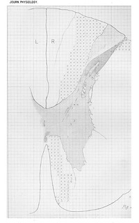
Fig. 14 in text:
'Passing upward at once without further description to the level of the 2nd thoracic nerve-root (Fig. 14) the following condition is discovered by the sections. A wide band of comparatively slightly degenerated tissue extends between the area of absolute degeneration and the posterior horn. If the whole area of the posterior column measures about 1050, the absolute degeneration in it measures 366. The area of absolute degeneratioin is wedge-shaped and extends a good half way toward the posterior commissure; and engages more than half (4/7) the free edge of the posterior column.' (278)
- No links match your filters. Clear Filters
-
Cited by
 C.S. Sherrington, 'Note on the Spinal portion of some Ascending Degenerations', Journal of Physiology 14 (4-5) (1893), pp. 255-302.
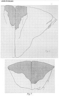
Description:Fig. 11 in text:
'An area of almost absolute degeneration skirts the posterior limb of the substantia gelatinosa; between this deep zone of degeneration and the great superficial area there is a zone of less severe degeneration, in which numbers of healthy nerve-fibres exist, both coarse and fine of various degrees of size; very few of the fibres are quite large, none of them so large as the fibres of the cerebellar tract; the majority are fine, averaging less than 2 µ. The area of absolute degeneration contains a scanty number of nerve-fibres still in process of degeneration, but the bulk of it is composed of cicatricial tissue in which numerous spaces represent the places previously occupied by nerve-fibres. The topography of the degeneration here can best be gathered from fig. 11.' (272)
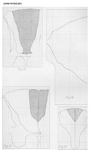
Figs. 15-16 in text:
'The posterior column which is much larger in area than at the last level examined contains some obvious scattered degeneration in its deepest part, that nearest to the grey matter. About 60-70 degenerating fibres can be found thLere in each section (Fig. 15). The area of absolute degeneration is less wide from right to left than it is lower down the cord. Its area is not really much less than at the level of the 8th cervical (231-254), but relatively to the whole posterior column instead of being a fifth part of it it is little more than a tenth.
At the level of the 2nd cervical nerve-root (Fig. 16) the posterior column of the cord although much larger than at the 2nd thoracic is not so large as at the 5th cervical. Yet the area of absolute degeneration in it is not only smaller absolutely than at the last mentioned level, but it is relatively smaller also, i.e. considerably less than one-tenth of the total column instead of somewhat more than one-tenth. Its edge is at least as sharp as previously. It is somewhat compressed laterally, so that it occupies less than a sixth of the free edge of the column. It is conterminous for the greater part of its side with a small septum. The area of the posterior column outside the degenerated area does not contain an obvious amount of scattered degeneration. There are indeed eight fibres in it that appear to be degenerated, but more I cannot detect; the position of these fibres is marked on the analysis map.' (280-281)
Fig. 16 in text:
'It is instructive to compare the configuration of the ascending degenerations at this level with the descending degeneration examined at the same level in the same species four months after a very large cortical lesion involving more than the whole "cord-area" of the cortex outside the gyrus fornicatus (Fig. 16 A). In the Macacque the crossed pyramidal tract extends outside its main mass into a peripheral zone described by France [note: 'Philos. Transacts. vol. B. 1889.'] and by myself [note: 'This Journal, March, 1889.']; in fcetus of the Macacque I find this peripheral sheet acquire myelin at the same epoch as does the pyramidal tract elsewhere; between this peripheral sheet (which in some specimens reaches halfway down the thoracic region) and the main body of the tract is an interval occupied almost exclusively by a coarse-fibred (megalomitous) tract, this is shown to degenerate (Fig. 16) upwards, and is the direct cerebellar. Although there is considerable overlapping of the territories of the ascending and descending degenerations the converse character of the general picture offered by the two degenerations is unmistakeable. The distinction between the ventral part of the lateral reticular formation and the part occupied by 'pyramid' fibres is sharp; in the foetus by paucity of myelin in the latter, in the adult by the ventral containing small fibres (micromitous), the latter fibres of very various sizes (poikilomitous).' (281-282)
Fig. 17 in text:
'The anatomy of the degeneration existing below the site of the lesion deserves some mention. The transverse lesion was at the 10th thoracic segment and involved the whole extent of the cord. A preparation taken below the level of origin of the 11th thoracic root about 17 mm. below the lesion is well comparable with a preparation from a level between the 9th and 8th roots because the anatomy of the normal cord is in the two places only slightly dissimilar. The antero-lateral column has a heavy degeneration throughout the greater portion of its dorsal half. The distribution of this is shown by the deeper shading in Fig. 17.' (282)
'A comparison is interesting between this descending degeneration two segments below a transverse spinal lesion and the descending degeneration of the pyramidal tract at the same level resulting from ablation of the whole "cord region" of the cortex (except Schäfer and France's gyrus fornicatus area). Not only is the area of descending degeneration (Fig. 19) in the lateral column much more extended in the case of the spinal lesion than in that of the cortical (Fig. 17 A) but within the area itself the density of the degeneration is much greater. Commingled therefore with cerebial fibres descending from the "cord-region" of the cortex are, as well as ascending spinal fibres, fibres which descend and are probably of spinal, some possibly of cerebellar (Marchi) [note: 'Rivista Sperim. di Freniatria, 1888.'] origin, all of them contained within the field appropriated by nomenclature to the crossed pyramidal tract [note: 'Sherrington. This Journal, 1885.'].' (282)
'There is another area of heavy degeneration in the most ventral portion of the anterior column, and along the margin of the ventral median fissure. A scattered degeneration of much slighter intensity involves the whole of the rest of the antero-lateral column, except for a narrow zone immediately abutting upon the grey substance. This narrow zone is narrowest at the free apex of the lateral horn and at the extreme ventral tip of the anterior horn; it is widest in the inlet of white matter bounded ventrally by the lateral horn dorsally by the base of the posterior horn, and it is fairly wide at the bottom of the anterior column. In each posterior colunmi a small oval patch of degeneration lies imbedded deeply, the mesial end nearly meeting the border of the grey matter, its other end directed toward the place of entrance of the posterior root (Fig. 17).' (283)


