- Creation
-
Creators (Definite): Sir Charles Scott Sherrington; The Cambridge Engraving CompanyDate: 1893
- Current Holder(s)
-
- No links match your filters. Clear Filters
-
Cited by
 C.S. Sherrington, 'Note on the Spinal portion of some Ascending Degenerations', Journal of Physiology 14 (4-5) (1893), pp. 255-302.
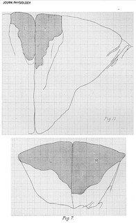
Description:Fig. 1 in text:
'Microscopical Examination. At the level of the lowest point of the scar there is (Pl. XIII., Fig. 1) a wedgeshaped area of degeneration with sclerosis at the outer free angle of the lateral division of the posterior column, involving nearly the whole width of the division of the column, and extending into the depth of the column as far as the apex of the substantia gelatinosa of the posterior horn. The sclerosis and the degeneration one millimeter higher is most dense in the portion abutting on the fissure between the lateral and posterior columns, and in a small part of that region no sound fibres at all are to be found.' (256)
Fig. 2 in text:
'One millimeter higher. The width of the area of degeneration and sclerosis has at the base of the lateral-posterior column become greater, but it still does not pass into the median-posterior column. The area extends through the entire depth of the lateral-posterior column. The part adjoining the fissure between the lateral and posterior column contains no healthy nerve-fibres. The scar is here attached for a slight extent to the subpial membrane of the cord in front of the fissure, and the most posterior edge of the cerebellar tract, where it lies against the entrance of the posterior root, is degenerated; a few healthy fibres existing among the degenerated. There is no obvious sclerosis here. One millimeter higher (Fig. 2). The width of the area of degeneration and sclerosis has at the base of the lateral-posterior column become greater, and reaches the outer edge of the median-posterior column. It passes through the entire depth of the lateral-posterior column. In the region abutting on the septum between the lateral and posterior column no healthy nerve-fibres are seen.' (256-257)
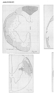
Fig. 3 in text:
'Just below the pyramidal decussation (Fig. 3). The degeneration in the most lateral part of the lateral posterior column is very severe; degeneration in that column occupies the whole width of its more superficial part, but becomes of slight intensity next to the septum dividing the column from the median posterior. The area of the whole right hand posterior column being taken as 415 (weight in lead foil), the portion of it containing signs of degeneration amounts to 267, and of this portion in an area of 57 the degeneration is very severe, approaching absolute.' (257)
Fig. 4 in text:
'At the level of the upper part of the pyramlidal decussation (Fig. 4) where the decussation of the fillet exists well marked, the area of degeneration in the right funiculus cuneatus in shape and extent differs little from that at the level just described. It has however a broader line of contact with the underlying grey matter at the base of what was the cornu posterius.' (258)


