- External URL
- Creation
-
Creator (Definite): Sir Charles Scott SherringtonDate: 1894
- Current Holder(s)
-
- No links match your filters. Clear Filters
-
Cites
 C.S. Sherrington, 'Note on the Spinal portion of some Ascending Degenerations', Journal of Physiology 14 (4-5) (1893), pp. 255-302.
Description:'The actual number of root-ganglion nerve-fibres in an ordinary muscular nerve-trunk is considerable. I have in not a few instances counted them, using the same method of counting as for the fibres of the spinal cord [note: 'This Journal, XIV. p. 287.' (224-225)
Sherrington's 1893 paper also highlights the anatomy of the posterior spinal root considered in detail in 'On the Anatomical Constitution':
'the above observations contribute an item toward illustration of an arrangement indicated by many researches that bear upon the afferent entries of the cord, namely, that a posterior spinal root establishes direct connections ('adjunctions' to judge by the results of the Golgi method) on its own side of the median plane not with one but with a great number of the chain of segments of the neural axis (e.g. afferent fibres from the leg plunging directly into grey matter of brachial spinal segments), whereas evidence of such direct relation of the fibres of a motor root to more than one or some very limited number of spinal segments is still wanting or at present even negatived.' (300)
-
Cites
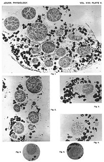
 Plate V, Journal of Physiology 17 (3-4) (1894). Figs. 1-6 from C.S. Sherrington, 'On the Anatomical Constitution of Nerves of Skeletal Muscles; with Remarks on Recurrent Fibres in the Ventral Spinal Nerve-root'.
Description:Explanation of plate V (figs. 1-6):
'Fig. 1. Spinal root-ganglion fibres in a large branch of the hamstrilng nerve to the medial hamstring muiscles (Monkey). Fifty-two days degeneration allowed after section of the 5th, 6th, 7th, 8th and 9th post-thoracic ventral and dorsal spinal roots, the latter cut proximal to their ganglia.
Fig. 2. Spinal root-ganglion fibres in a branch to tibialis posticus and flexor digitorum longus (Cat), 6th, 7th, 8th and 9th post-thoracic ventral aind dorsal spinal roots severed, the latter proximal to ganglia. Forty-nine days degeneration allowed.
Fig. 3. Spinal root ganglion fibres in a branch from hamstring nerve to biceps femoris; fifty-eight days after section of 6th, 7th, 8th and 9th postthoracic venltral and dorsal spinal roots, the latter proximal to ganglia. A small nerve-twig to left hand consists only of large root-ganglion fibres.
Fig. 4. Twig from "kneejerk" nerve (vastus medialis). All except root-ganglion fibres removed by forty-two days degeneration. A smaller twig, shows very little sclerosis and contains large and small myelinate fibres.
Fig. 5. Twig to femoralis, normal (Monkey).
Fig. 6. Corresponding opposite nerve from same monkey; spinal rootganglion fibres alone left. 3rd, 4th, 5th and 6th post-thoracic ventral and dorsal spinal roots cut, the latter proximal to ganglia. Fifty-four days degeneration allowed.' (256-257)
-
Cites
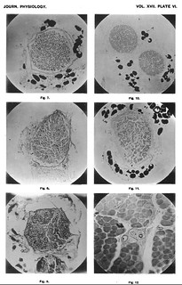
 Plate VI, Journal of Physiology 17 (3-4) (1894). Figs. 7-12 from C.S. Sherrington, 'On the Anatomical Constitution of Nerves of Skeletal Muscles; with Remarks on Recurrent Fibres in the Ventral Spinal Nerve-root'.
Description:Figs. 7-12 in text:
'Fig. 7. Twig to seminembranosus, normal (Cat).
Fig. 8. Corresponding from opposite side, on which the 6th, 7th, 8th and 9th post-thoracic spinal ganglia and ventral roots had been excised thirty-four days. No myelinate fibres left.
Fig. 9. Corresponding twig from another cat of about the same size as preceding, in which 6th, 7th, 8th and 9th post-thoracic ventral and dorsal spinal roots had been cut, the latter proximal to ganglia. Thirty-four days degeneration allowed.
Fig. 10. Nerve to vastus medialis and adjoining part of femoralis (Monkey). Knee-jerk nerve. Normal.
Fig. 11. Corresponding nerve of opposite side, the 3rd, 4th, 5th and 6th post-thoracic ventral and dorsal spinal roots having been cut, the latter proximal to ganglion. Thirty-five days degeneration allowed.
Fig. 12. Flexor digitorurm brevis muscle (Cat); root-ganglion fibres only remaining. 6th, 7th, 8th, and 9th post-thoracic ventral and dorsal spinal roots severed, the latter proximal to ganglia. Thirty-five days allowed for degeneration. Small intra-muscular nerve-twigs in cross section; from the smaller of these (purely motor) all myelinate fibres have disappeared, from the larger all but two (one 16µ, one 2µ). Near the smaller twig a small artery. Further to the right and above is the commencement of a "muscle spindle;" into the capsule of this the large fibre seen in middle of figure was traceable in the series of sections made.' (257)
Figs. 5, 7 and 10-11 in text:
'for the present purpose it is necessary to be certain that the nerve-trunk selected is actually destined for muscle. In the term "muscle" I here include its connective-tissue accessories of tendon, perimysium, &c. It is necessary therefore to select nerve-trunks which are actually plunging into the fleshy parts of ample muscles. Now, when corresponding nerve-twigs of this kind are taken from the right and left limbs respectively it is found that on microscopic examination they frequently do not prove so symmetrical as one would wish for carrying out their comparison (cf. figs. 10, 11, plate VI.). Thus:
[3 tables comparing left/right distributions of nerve fibres here]
I have endeavoured to overcome this discrepancy, which though sometimes trifling is more frequently wide, by gauging the number of fibres that must originally have been present in a nerve-branch by measurement of its transverse sectional area. In normal muscullar nerves the fibres in the nerve-bundles are regularly and compactly arranged (cf figs. 5, 7, 10, plate VI.). A given considerable area of cross section of nerve-bundle contains, if the same procedure in fixing and dehydration has been used, in the same individual not far from the same absolute number of fibres in corresponding nerves of the right and left sides.' (226-227)
-
Cites
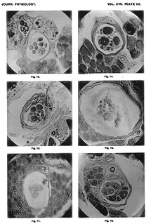
 Plate VII, Journal of Physiology 17 (3-4) (1894). Figs. 13-18 from C.S. Sherrington, 'On the Anatomical Constitution of Nerves of Skeletal Muscles; with Remarks on Recurrent Fibres in the Ventral Spinal Nerve-root'.
Description:Explanation of Plate VII (figs. 13-18):
'Fig. 13. Gracilis muscle, Monkey; root-ganglion fibres only remaining. Muscle-spindle containing eight muscle-fibres: near it a "spindle nerve" containing five large myelinate fibres; also a motor nerve from which all nyelinate fibres have disappeared. The periaxial space is well seen.
Fig. 14. Adductor hallucis muscle (Monkey). 6th, 7th, 8th and 9th post-thoracic ventral and dorsal spinal roots severed, the latter proximal to ganglia. Fifty days degeneration allowed. A compound "muscle-spindle" in cross section near one of its poles. Three large muscle-fibres and five small inside the spindle. A large thinly myelinate nerve-fibre is seen cut twice, obliquely, in the capsule of the organ.
Fig. 15. Vastus medialis muscle (Cat). 4th, 5th, 6th and 7th postthoracic ventral and dorsal spinal roots severed, the latter proximal to ganglia. Sixty days degeneration allowed. Muscle-spindle through its proximal polar region containing three large and six small muscle fibres. A large root-ganglion fibre lies in the outer part of the capsule of the organ, a smaller myeliniate fibre lies close to the muscle-fibres in the core of the organ.
Fig. 16. Biceps femoris (Cat). 6th, 7th, 8th and 9th post-thoracic ventral and dorsal spinal roots cut, latter proximal to ganglion. 106 days degeneration allowed. A "muscle-spindle" in cross section through its equatorial region. Three thinly myelinate nerve-fibres obliquely cut in the periaxial space. Nine muscle-fibres and two non-myelinate terminal nerve fibres seen in core. The delicate tissue in the periaxial lymph space is visible.
Fig. 17. Semimembranosus muscle (Monkey). 6th, 7th, 8th and 9th post-thoracic ventral and dorsal spinal roots severed, latter proximal to ganglia. Fifty days degeneration allowed. A "muscle-spindle" in cross section through the equatorial region. Two myelinate fibres cut obliquely in the thickness of the capsule of the spindle; three cut fairly transversely just within the periaxial lymph-space. Six muscle-fibres and two non-myelinate nerve-fibres cut transversely within the core of the organ.
Fig. 18. Interosseous plantar muscle (Cat). Compound "muscle-spindle" in distal polar region not far from junction with equatorial region. One division of the spindle contains eight muscle-fibres, the other nine muscle fibres. In the smaller division a myelinate fibre is seen in transverse section, in the larger a myelinate fibre curves across from the centre to the periphery of the intrafusal bundle. At one side of the spindle lies a large solitary "spindle" nerve-fibre enclosed in a well-developed loosely-fitting sheath of Henle.' (257-258)
Fig. 13 in text:
'besides isolated nerve-fibres attaining the spindles, not infreqtient are small nerve twigs conisisting of 3-6 large spinal-ganglion fibres all bound for one and the same muscle-spindle and entering it at one point, or two closely adjacent points. These twigs are pure spindle-nerves. Such spindle-nerves form one class of the purely afferent intramuscular nerve-trunks above described. One such is figured in Fig. 13, Plate VII.' (244)
Figs 16-17 in text:
'These septa, as also the membranes of the capsule proper, are studded with sparse oval nuclei like those upon the coats of a Pacinian corpuscle. The open work of delicate septa webbing across the periaxial lymph space fuses on one side with the inner face of the inmost sheet of the corpuscle proper, on the other with a thin more or less perfect layer of connective tissue which immediately invests the axial core of the spinidle. This investment I would term the axial sheath. It is fairly well shown in figs. 16 and 17, Plate VII. It is richly nucleate and supports as well as invests the neuro-muscular contents of the organ. In a compound spindle each intrafusal muscle-bundle retains its own axial sheath, even where the capsules of the component spindles are completely merged one in another.' (240-241)
Fig. 16 in text:
'In the equatorial region the intrafusal muscle-fibre often becomes somewhat smaller in diameter and is nearly always circular in section. Its surface zone soon gets thickly encrusted with or almost completely occupied by a sheet of nuclei. Whether these nuclei are strictly part of the muscle-fibre proper is not clear to me. The nuclei are spherical or slightly oval, are clear, and measure about 6µ diameter. Cross-sections reveal beneath the nuclear sheet a thin tuibular layer which is fibrillated. This tubular layer itself invests a central core (4µ-5µ) of hyaline substance, which runs rodlike along the axis of the intrafusal fibre in this region (Fig. 16). The cross-section of the fibre thus often displays a nearly complete zone of four to six nuclei around a hyaline centre. The striation is obscured by the nuclei clothing the face of the attenuated fibre, but where seen in the adjoining regions the striation is very marked, and coarser than in the ordinary muscle-fibres outside the spindle.' (242)
'The equatorial region usually occupies somewhat less than one-third of the entire length of the spindle (Fig. 16, Plate VII.). On emerging from this region the intrafusal fibres quickly regain the character they possess in the proximal polar region. The number of nuclei in them becomes less as the distal pole of the spindle is approached.' (243)
-
Cited by
 Charles S. Sherrington and 'Mechanical Objectivity'.
Description:'Rather than present lavish illustrations of his nerve investigations, as had Gaskell (fig. 1), Sherrington supplied tables of response measurements, graphs of reaction-intensities, and introduced grids with which he might reduce the complexity of nervous anatomy (fig. 2). His study addressing the pioneering histological work of Angelo Ruffini replaced the latter's all-encompassing hand-drawn delineations (fig. 3) with a series of microphotographs (fig. 4).'
-
Cited by
 Langley to Sherrington - 8 May 1922 (I-2-135 (iv))
Description:'I have just received your letter. 'Mayitol' & the specimens will no doubt come in the next post. But I am writing at once since it occurs to me you may have kept specimens or nerves of your two sets of 1894 in which you received the lower lumbar ganglia omitting the posterior roots (Journal, 17, p. 223). I fear neither nerves nor specimens can have lasted till now, but if they have I should like to look at them. The point is a detail of no particular importance, but I am a little doubtful whether all the postganglionic fibres lose their myelin in the cat soon after joining the spinal nerves.'
-
Cited by
 T. Quick, 'Disciplining Physiological Psychology: Cinematographs as Epistemic Devices, 1897-1922', Science in Context 30 (4), pp. 423-474.
Description:'the mode in which Sherrington presented his work owed as much to the formulae, tables and calculations of the Cavendish physicists as it did to the anatomic accuracy of his Cambridge mentors. Rather than present lavish illustrations of his nerve investigations, as had Gaskell, Sherrington supplied tables of response measurements, graphs of reaction-intensities, and introduced grids with which he might reduce the complexity of nervous anatomy (fig. 1). His study addressing the pioneering histological work of Angelo Ruffini replaced the latter's all-encompassing hand-drawn delineations with a series of microphotographs (fig. 2) (Sherrington 1893; Sherrington 1894).'


