- Creation
-
Creators (Definite): Sir Charles Scott Sherrington; Albert Frank Stanley KentDate: 1894
- Current Holder(s)
-
- No links match your filters. Clear Filters
-
Cited by
 C.S. Sherrington, 'On the Anatomical Constitution of Nerves of Skeletal Muscles; with Remarks on Recurrent Fibres in the Ventral Spinal Nerve-root', Journal of Physiology 17 (3-4) (1894), pp. 210-258.
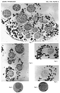
Description:Figs. 7-12 in text:
'Fig. 7. Twig to seminembranosus, normal (Cat).
Fig. 8. Corresponding from opposite side, on which the 6th, 7th, 8th and 9th post-thoracic spinal ganglia and ventral roots had been excised thirty-four days. No myelinate fibres left.
Fig. 9. Corresponding twig from another cat of about the same size as preceding, in which 6th, 7th, 8th and 9th post-thoracic ventral and dorsal spinal roots had been cut, the latter proximal to ganglia. Thirty-four days degeneration allowed.
Fig. 10. Nerve to vastus medialis and adjoining part of femoralis (Monkey). Knee-jerk nerve. Normal.
Fig. 11. Corresponding nerve of opposite side, the 3rd, 4th, 5th and 6th post-thoracic ventral and dorsal spinal roots having been cut, the latter proximal to ganglion. Thirty-five days degeneration allowed.
Fig. 12. Flexor digitorurm brevis muscle (Cat); root-ganglion fibres only remaining. 6th, 7th, 8th, and 9th post-thoracic ventral and dorsal spinal roots severed, the latter proximal to ganglia. Thirty-five days allowed for degeneration. Small intra-muscular nerve-twigs in cross section; from the smaller of these (purely motor) all myelinate fibres have disappeared, from the larger all but two (one 16µ, one 2µ). Near the smaller twig a small artery. Further to the right and above is the commencement of a "muscle spindle;" into the capsule of this the large fibre seen in middle of figure was traceable in the series of sections made.' (257)
Figs. 5, 7 and 10-11 in text:
'for the present purpose it is necessary to be certain that the nerve-trunk selected is actually destined for muscle. In the term "muscle" I here include its connective-tissue accessories of tendon, perimysium, &c. It is necessary therefore to select nerve-trunks which are actually plunging into the fleshy parts of ample muscles. Now, when corresponding nerve-twigs of this kind are taken from the right and left limbs respectively it is found that on microscopic examination they frequently do not prove so symmetrical as one would wish for carrying out their comparison (cf. figs. 10, 11, plate VI.). Thus:
[3 tables comparing left/right distributions of nerve fibres here]
I have endeavoured to overcome this discrepancy, which though sometimes trifling is more frequently wide, by gauging the number of fibres that must originally have been present in a nerve-branch by measurement of its transverse sectional area. In normal muscullar nerves the fibres in the nerve-bundles are regularly and compactly arranged (cf figs. 5, 7, 10, plate VI.). A given considerable area of cross section of nerve-bundle contains, if the same procedure in fixing and dehydration has been used, in the same individual not far from the same absolute number of fibres in corresponding nerves of the right and left sides.' (226-227)


