 Material relating to Charles Sherrington's work on ocular physiology and binocular vision
 Material relating to Charles Sherrington's work on ocular physiology and binocular vision

- No links match your filters. Clear Filters
-
Related to
 'Proceedings of the Physiological Society', 7 (3) (1886), pp. xiii-xvii.
Description:'Mr SHERRINGTON exhibited a rabbit, in which he had placed a ligature round the optic nerve of the right side nine weeks previously. The operation had been performed witth antiseptic precautions, and the ligature used was of catgut, about a millimeter thick. This ligature was tied as tightly as possible.
On the evening of the day of operation the retinal vessels as conmpared with those of the healthy side were reduced to very fine streaks, perhaps a tenth of their previous diameter; no pulsation could be produced in them by compression, although that was easily done for the other eye.
The tension of the eyeball instead of being increased, as stated for rabbits by Prof. Stilling, was about -1.
Observation of the fundus was impossible for the next fortnight, because of opacity of the media, but duiring the whole of that period the tension on the ligatured side was never so great as on the healthy side.
At the present time, as was demonstrated, the tension was still lower than in the healthy eyeball. Moreover the retinal circulation has become reestablished, although the vessels, especially the arteries, are smaller than in the opposite side.
Pulsation becomes evident in these vessels on pressure being applied to the eyeball, just as on the uninjuired fundus of the left side.
The right eye is however completely blind. The iris does not react to light. Otherwise the eye to cursory examination would appear normal. Closer observation detects merely a slight nebula, the remnant of a keratitis that occurred in the first fortnight following the operation; and the refraction has become myopic, and astigmatic, being - 2 D as measured on the vertical vessels of the disc, and - 3.5 D as measured on the horizontal.
At the optic disc itself papillitis is passing, into atrophy.'
-
Related to
 'Proceedings of the Physiological Society', Journal of Physiology 20 (Supplement) (1896), pp. xi-xxii.
Description:'Influence of simultaneous contrast on "flicker" of Visual Sensation - By C. S. SHERRINGTON.
It was demonstrated upon a number of rotating discs that the "flickering" of sectors of a disc turning at speeds insufficient to give perfectly fused sensation is much influenced by "simultaneous contrast" acting upon those sectors. Contrast could thus be made to lower or heighten the speed of rotation requisite to cause steady combination of two colours. The amount of lowering or heightening obtainable was very considerable indeed, raising the minimum frequency of intermittence allowing fusion for the same couple of physical colours from e.g. 22 to 34 per second, or again from e.g. 35 to 51 per second. It was pointed out that the method showed it possible to dismiss from consciousness all difference between the different backgrounds causing contrast to appear in stripes laid on them and yet retain consciousness of a difference between the stripes. It was also pointed out that the method gives a ready and accurate means of obtaining measurement of quantity of contrast-effect, i.e. in terms of frequency of intermittence, that is in the same terms as of the "flicker" method of colour photometry introduced by Rood.' (xviii-xix)
[nb cites 'Rood, American Journal of Science, XLVI. p. 173.']
-
Related to
 C.S. Sherrington 'Experimental Note on Two Movements of the Eye', Journal of Physiology 17 (1-2) (1894), pp. 27-29.
Description:Concerns the stimulation of the cerebral cortex in monkeys in order to obtain ocular movement.
Severance of the 3rd and 4th cranial nerve of one side of the eye facilitates excitation of the cortex that 'produces conjugate movement of both eyes towards the opposite side, i.e. from left toward right, the left eye travelling however onbly so far as the median line.' (27)
'As regards the question, 'Where does the action of arrest take place, is it cortical or subcortical?... at least in many cases... the cortex is not necessary for the reaction.' (27) - Outlines series of ablation procedutres to demonstrate this. (27-28)
Further notes on experiments described in previous paper. (28)
Concludes that 'it seems clear that this inhibition which can be elicited by experimental excitation of the grey cortex and of the underlying white matter is fully and habitually excercised in volitional eye movements...
For the eyeball under simple relaxation of a rectus muscle to revert from a direction of deviation back to the primary position, its setting in the orbit must be such that the tensions of its connections, apart from active contraction or nervous tonus in its extrinsic muscles, are in equilibrium only when the globe is in the primary position.' (29)
Notes that observations of eyelid movements 'after intercranial section of the IIIrd nerve': 'Under volitional [non-stimulated] action the opening of the right eye was never once seen to be accompanied by any movement at all in the lids of the left. In blinking the opening after momentary closure was as rapid on the left side as on the right. That is to say there was no evidence of any cooperative contraction of the antagonist muscles under volition any more than under experimental cortical excitation or during epilepsy.' (29)
-
Related to
 C.S. Sherrington, 'Further Note on the Sensory Nerves of the Eye-Muscles', Proceedings of the Royal Society of London 64 (1898-1899), pp. 120-121.
Description:'"Further Note on the Sensory Nerves of the Eye-Muscles." By C. S. SHERRINGTON, M.A., M.D., F.R.S., University College, Liverpool. Received September 21, Read November 17, 1898.
In a communication on the sensory nerves of muscles laid before the Society last year, the question previously raised as to the existence of afferent nerve fibres in the so-called "motor" cranial nerves for the muscles of the eye was advanced to the following position. Myelinated nerve-fibres from the IIIrd cranial and IVth cranial nerve-roots were traced into the tendons of the recti and oblique muscles. Since then control observations have been made. The ophthalmic division of the Vth cranial has been severed at its origin, and with it the Vlth cranial trunk. This has been done in the monkey. The condition of the nerve branches going to and lying within the muscles and tendons has been examined after an interval of twelve to fourteen days. Those of the external rectus contained nothing but degenerate nerve-fibres, save for a few fine myelinate fibres, probably from the ciliary ganglion. Those of all of the other eye-muscles contained exclusively healthy nerve-fibres. The sensorial musculo-tendinous organs of the eye-muscles are, therefore, not innervated by the ophthalmic division of the tri-geminus. On the other hand, the nerve-fibres of the external rectus muscle behave, after severance of the VIth cranial nerve, in the same way as my previous papers showed those of the other eye muscles do after section of the IIIrd and IVth nerves.A contribution towards the physiological inquiry into the matter has been made as follows, and has given a clear reply. The conjunctives, both palpebral and ocular, and the corneas of both eyes have been rendered deeply anaesthetic to cold, warmth, touch, and pain by liberal applications of cocaine. Then in a completely dark room the power to direct the gaze with accuracy in any required direction has been tested. The person under examination is seated, with the head securely fixed, in front of a screen. One of his hands carries a marker. The hand is moved by an assistant, and is made to mark the screen at some one point; it is then passively replaced. The person under observation during this time keeps the eyes open in the primary position or sits with them closed. He is then required to direct his gaze to the spot marked on the screen. The light is then switched on, and the point to which the gaze is turned is noted. The power to direct the gaze under these circumstances has been found to remain good. If for co-ordinate execution and ability to perform the delicate adjustments for training the eyeballs correctly in any desired position the exercise of peripheral apparatus of muscular sense is required, the only possible channels under the above conditions would seem to be deep branches of the Vth nerve or the IIIrd, IVth, and VIth so-called " motor " nerves themselves. As previously stated, the former are by both my earlier and later degeneration experiments excluded. The latter, therefore, are the only ones remaining, for the superficial branches of the Vth and the retinae are put out of action by the conditions of experiment.
I am indebted to Mr. E. E. Laslett for carrying out the observations with me. Details regarding the methods employed and the results obtained will be given in a completer paper written in conjunction with him.
[NB, Refs to C. S. Sherrington, ' Roy. Soc. Proc.,' vol. 61, p. 247, February, 1897. and C. S. Sherrington, ' Physiol. Soc. Proc.,' No. 3, June, 1894.] -
Related to
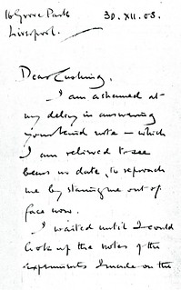
 Sherrington to Cushing - 30 December 1908 (WCG 32.16)
Description:'I waited until I could look up the notes of the experiments I made on the eye-muscle nerves. I have not published anything further than the reference in Roy. Soc. Proc. of '97. I think that was [the] date. The notes made at the time show that the III and IV nerves were cut in 5 monkeys, & cut at emergergence from brain. The myelinate nerve-fibres in the muscles were looked at, at dates from 12 to 36 days; it was found that the numerous (e.g. 250) myelinate nerve fibres which reach the insertion end of the muscle in every case showed heavy degenreration. A few (e.g. 30 or so) of these were not degenerate: these were all of small calibre. We concluded these must come from Vth or from ciliary or sympathetic. On cutting Vth (this was done only in one case) distal to ganglion no degenerated myelinate fibre was found in any of the eye-muscles. I concluded that the few undegererated fibres in muscle tendon must come from synap. or from ciliary ganglion.
Now the myelinate nerve fibres at insertion end of muscle must be afferent one imagines, many of them hook down into tendon & then return. There I think can hardly be motor
[sketch at bottom of page: 'myelinated nerve fibres']
for one finds their ends by methylene blue method, as figured in [G. Carl] Huber's paper. Besides I could find no plates at ends of muscle.
Your experiments interest me therefore very much. Our results seem as regards IIIrd n. to be squarely opposed. My wounds healed all night & the ext. rectus muscle nerves showed no trace of degen.
What has always kept me back from publishing my results more fully has been the failure to be able to suggest what cells can give origin to afferent fibres in the 3rd nerve. The so called degenerate ganglion does not seem to, for as far as I could see the fibres went clean through it. Are there any sensory type cells in the IIIrd nerve nucleus in the side of & "below iter[?] a IIIiv[?] and IVam[?] ventriculum"? To ascertain this I cut a number of sections of monkey's isthmus region & could not see such cells. I also went to Louvain and looked through [related to Wautier Franscois?] Van Genechten's series of IIIrd nucleus specimens & could not satisfy myself of any clearly sensory cells in IIIrd nucleus: nor could he (I put question to him).
The point is interesting: I will try for my own satisfaction to repeat my observations & see if I get same results. There is some 'Kunstfehler' somewhere, & I suspect it must by of my commission, & not yours. But I do not see where it lies. H.K. Anderson had the ciliary ganglion in one of my specimens & he detected nothing wrong in my operation.'


