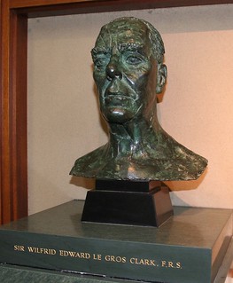
Sir Wilfred Le Gros Clark conducted a number of studies on the neuroanatomy of the visual system, culminating in his 1942 review paper on the visual centres of the brain, Le Gros Clark, W. E. 1942. The visual centres of the brain and their connexions. Physiological Reviews. 22, 205-232.
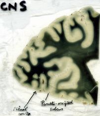
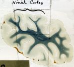
Slides 7, 8, 9, 10, 13 and 14 in drawer 7 exhibit features of Le Gros Clark’s studies on the anatomy of the visual cortex of the rhesus monkey. Slides 7, 8, 9 and 10 show general features of the visual cortex of the rhesus monkey, including the thin medullary lamina, the white line of Gennari (slides 7 and 8), the deep calcarine sulcus in which the visual cortex lies (slide 9) and the layers of the cortex (slide 10).
Slide D13 in drawer 7 appears to be the source of Figure 1 (p 371) in Le Gros Clark (1942), Le Gros Clark, W. E. 1942. The cells of Meynert in the visual cortex of the monkey. Journal of Anatomy, 76 (4), 369-376.1. The composite drawing in this figure is a high magnification of cells of the visual cortex of the rhesus monkey, allowing the identification of two cells of Meynert. From this study Le Gros Clark (1942) concluded (p 325) that the cells of Meynert are situated mainly in lamina VI of the visual cortex and can be characterised by their basal dendrites. It was concluded that the axons of the Meynert cells contribute to the cortico-mesencephalic connexions.
A more recent study on Meynert cells in the visual cortex of rhesus monkeys, Chan-Palay, V., Palay, S. L., Billings-Gagliardi, S. M. 1974. Meynert cells in the primate visual cortex. Journal of Neurocytology, 3, 631-658, describes large Meynert cells in layer V of the striate cortex. These cells are argyrophilic and have a large number of neurofilaments in their dendrites and perikarya. It was found that each Meynert cell has an apical and many large basal dendrites. In layer II, the dendrites bursts into a large number of branches. Le Gros Clark and Sunderland (1939) found Meynert cells mostly in lamina VI and sometimes in the boundary between laminae V and VI, which corresponds quite well with the results from this study.
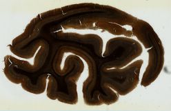
Slide D14 in drawer 7 appears to correspond to Figure 2B (p 571) in Le Gros Clark, W. E. and Sunderland, S. 1939. Structural changes in the isolated visual cortex. Journal of Anatomy, 73 (3), 563-574.3. In their attempts to analyse the functional significance of the laminar organisation of the cerebral cortex Le Gros Clark and Sunderland (1939) examined changes in the cells of the visual cortex of the rhesus monkey after isolation of small areas of visual cortex using knife cuts. This slide shows the discrete fasciculi of radial fibres running vertically through the lower levels of the cortex. The slide also shows fibres of varying size which are not collected in discrete bundles and other fibres that are in a closely meshed felt-work forming the stria of Gennari.
From this study the authors concluded that the isolated cortex showed a slight reduction of cells in all the lamina. Most of the large stellate cells of lamina IV and many of the large solitary cells of Meynert in lamina VI atrophied and disappeared. In the isolated cortex there was a reduction of the number of individual fibres, while the number of fasciculi which they compose, remained unchanged. It is possible that these radial fibres are derived from other parts of the visual cortex. The network of obliquely disposed fibres in the deeper levels of the cortex mostly disappears. It is suggested that these fibres are projection fibres derived from subcortical centres. The stria of Gennari appeared to remain unchanged in width and density.

- No links match your filters. Clear Filters
-


-
-
_CROPPED/TileGroup0/0-0-0.jpg)
-
_CROPPED/TileGroup0/0-0-0.jpg)
-
-

-