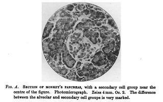
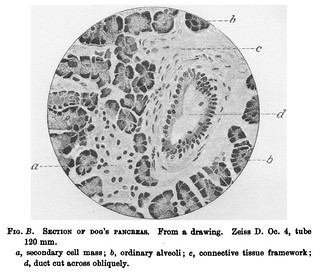
Caption:
'Fig. D. Section of Pancreas of Guinea-Pig. In the centre of the figure is a large multo-nucleated secondary cell group surrounded by the ordinary alveoli. Photomicrograph. Zeiss 2 mm. Oc. 2.' (356)
In text:
'The pancreas of the guinea-pig (fig. D) is characterised by the possession of particularly large and clear cells and large spherical nuclei, and by the fact that the stainiing reaction is by no means so definite as in those glands hitherto mentioned, and the same remark applies to the pancreas of the seal.' (354-355)
'In some cases the masses, more or less large and distinctly surrounded by a capsule of fine fibrous tissue, are not differentiated into distinct cells, but the nuelei are very numerous and stain deeply. Podvysotsky, who gave a description of the structure of the pancreas in 1884, evidently had observed only this type of secondary cell group, when he wrote, as he described the secondary collection of such cells as like lymphoid follicles. This type has certainly somewhat that appearance, but, as the author says, the collection of cells has in reality nothing in common with lymphoid elements. An exceedingly good example of this type is seen in the pancreas of the guinea-pig. In the photograph of this animal's pancreas (fig. D), the mass looks very like either a lymph follicle or a Malpighian corpuscle of the kidney uninjected.' (357)

- No links match your filters. Clear Filters
-


-
-

-
-

