- Creation
-
Creator (Definite): William Barnett WarringtonDate: 1899
- Current Holder(s)
-
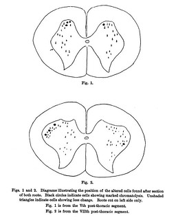
Caption:
'Diagram indicating position of altered cells after complete transection of the cord. Degeneration of white matter shown on one side. Sketch taken from midlumbar region. Black circles indicate cells showing chromatolysis. Unshaded triangles normal cells.' (474)
Fig. 3 in text:
'Exp. 1. Cat. Complete transection of the cord at the level of Ist lumbar segment; killed on the 15th day.
Series of sections cut in the several segments as low down as the VlIlth post-thoracic. The cord was found to be in good condition and quite free from inflammatory complications.
Sections were examined from below the site of the lesion to the lower part of the IInd lumbar segment.
In the cat Clarke's Column is well marked in this position and many of the cells showed undoubted chromatolysis.
The cord at the region of the enlargement was quite normal and no altered cells could be found.
In the middle of the lumbar region however a considerable number of the cells were in a distinct state of chromatolysis, but the large majority were unaffected. The situation of the altered cells was chiefly in the posterior part of the grey matter as shown in Fig. 3.
The condition of the individual cells did not differ materially from that described as resulting after other forms of physiological interference. The blue chromatic granules were disintegrated and the nucleus was often situated peripherally.' (474-475)

- No links match your filters. Clear Filters
-


-