- Creation
-
Creator (Definite): William Barnett WarringtonDate: 1899
- Current Holder(s)
-
Caption:
'Figs. 1 and 2. Diagrams illustrating the position of the altered cells found after section of both roots. Black circles indicate cells showing marked chromatolysis. Unshaded triangles indicate cells showing less change. Roots cut on left side only. Fig. 1 is from the Vth post-thoracic segment. Fig. 2 is from the VIIth post-thoracic segment.' (467)
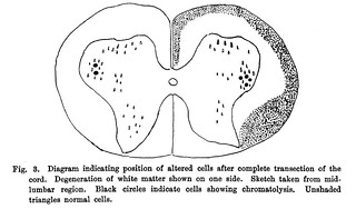
Fig. in text:
'All the larger cells on the side of the lesion showed changes. The majority were only comparatively slightly affected to the extent seen after section of an anterior root alone, others however showed a much more marked chromatolysis, the change resembling that found after section of the afferent roots.
In the Vth segment the anterior group was thus specially picked out, and in the VIth and VIIth the postero-lateral (Figs. 1 and 2).
This group is thus again brought into prominence on account of the ease with which its cells undergo chromatolysis, and the experiment affords further explanation of the results obtained in old standing amputations, where of course both afferent and efferent fibres are cut and yet the ultimate damage in the cord is nearly limited to this region of cells.' (466-467)

- No links match your filters. Clear Filters
-


-