Le Gros Clark, W. E. 1940. The nervous and vascular relations of the pineal gland. Journal of Anatomy, 74 (4), 471-492.
During the late 1930’s, neuroanatomists found that the pituitary gland (hypophysis) had both neural and vascular connections with the hypothalamus. In a paper published in 1940, Le Gros Clark investigated the neural and vascular connections of the pineal gland in the brain of the rhesus monkey and humans.
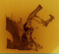
Slide E02 in drawer 9 appears to correspond to Figure 1 (p 476) of Le Gros Clark (1940). This slide shows the vascular supply of the pineal gland of the rhesus monkey from the great cerebral vein (vena magna cerebri) and a large amount of dark brown pigment in the melanocytes in the surrounding parenchyma.
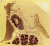
Slide E05 in drawer 9 appears to correspond to Figures 7 and 8 (p 484 and 485) of Le Gros Clark (1940). This shows the neural and vascular connections of the pineal gland of the human brain. Serial sections were prepared from a block removed from a human brain, showing the pineal gland and the attachments to the mid-brain, epithalamus, cerebellum, vena magna cerebri (great cerebral vein) and the straight sinus. This block of brain was embedded in cellodin following treatment with 5% nitric acid to decalcify the calcareous deposits. The tissue was cut at 30 and 150 µ and stained with haemotoxylin and eosin. These sections show that the gland has several calcareous masses. The nervus conarii (pineal nerve) travels from the tip of the pineal gland along the great cerebral vein then enters the base of the arachnoid granulation and finally enters the straight sinus. The large arachnoid granulation projects from the surface of the upper cerebellum to the opening of the straight sinus and contains several blood sinuses, which appear partly empty and collapsed. However, the granulation seems to elevate the floor of the anterior end of the straight sinus and narrows the opening of the vein of Galen. Because of its location, above and behind the pineal gland, the granulation can be described as the “suprapineal arachnoid body”.
Slide E06 in drawer 9 appears to correspond to Plate I, Figure 1 (p 493) of Le Gros Clark (1940). It shows the nervus conarii (pineal nerve) emerging from the pineal gland of the rhesus monkey and passing along the vena magna cerebri (great cerebral vein). During its course to the straight sinus, it breaks up into a plexus arrangement of fasciculi related to fine blood vessels running down to enter the tip of the pineal gland. The nerve is bounded in in a connective tissue sheath and resembles a peripheral nerve.
Slide E07 in drawer 9 appears to correspond to Plate I, Figure 2 (p 493) in Le Gros Clark (1940). It shows nerve cells that are mainly collected in the centre of the distal half of the pineal gland of the rhesus monkey where they are embedded in a mass of neuropil, forming the central core of the gland. The vascularity of this core can be seen as the clear areas between the intertwined nerve fibres.
From this research, Le Gros Clark (1940) concluded (p 490-491) that the pineal gland of the rhesus monkey contains a central core of neuropil containing a great number of large ganglion cells with branching processes. The central core is perfused by a rich capillary plexus. Branches of chorioidal arteries are accompanied by perivascular plexuses which penetrate the pineal gland from its lateral aspect. A chorioidal vessel extends into the straight sinus and this vessel has recurrent twigs descending into the apex of the pineal gland. A conspicuous fasciculus can be traced back to the apex of the pineal gland where it emerges as the nervus conarii (pineal nerve). This nerve can be traced from the central core of neuropil in the pineal gland into the wall of the straight sinus, where it branches in a plexiform manner. Some fibres of the posterior commissure terminate in relation to the ependymal epithelium lining the posterior wall of the pineal recess where they show regular beading and small end-bulbs. Some of his sections show a globular skein of nerve fibres in the pial tissue between the straight sinus and the pineal gland, which resembles a sensory nerve-ending.
From his research on the human pineal gland, Le Gros Clark (1940) concluded (p 491) that the nervus conarii (pineal nerve) emerges from the tip of the gland and runs an uninterrupted and unbranched course to reach the dura mater of the tentorium cerebelli. From there it runs back in the floor of the straight sinus in a subendothelial position. The formation of an arachnoid was described in relation to the floor of the straight sinus at the position where the great vein of Galen opens into it. The structure resembles an unusual, large arachnoid granulation, but its stroma consists of dense pial tissue containing a sinusoidal plexus of blood vessels and large blood sinuses. Because of its structure and location it has been termed the suprapineal arachnoid body. It is suggested that this body might provide a ball-valve mechanism in which it regulates the venous return from the great vein of Galen. In two specimens, the nervus conarii (pineal nerve) seems to pass through the suprapineal body and the blood sinuses on its way to the dural floor.

- No links match your filters. Clear Filters
-


-
-
-
-