Information
- Tags
Description
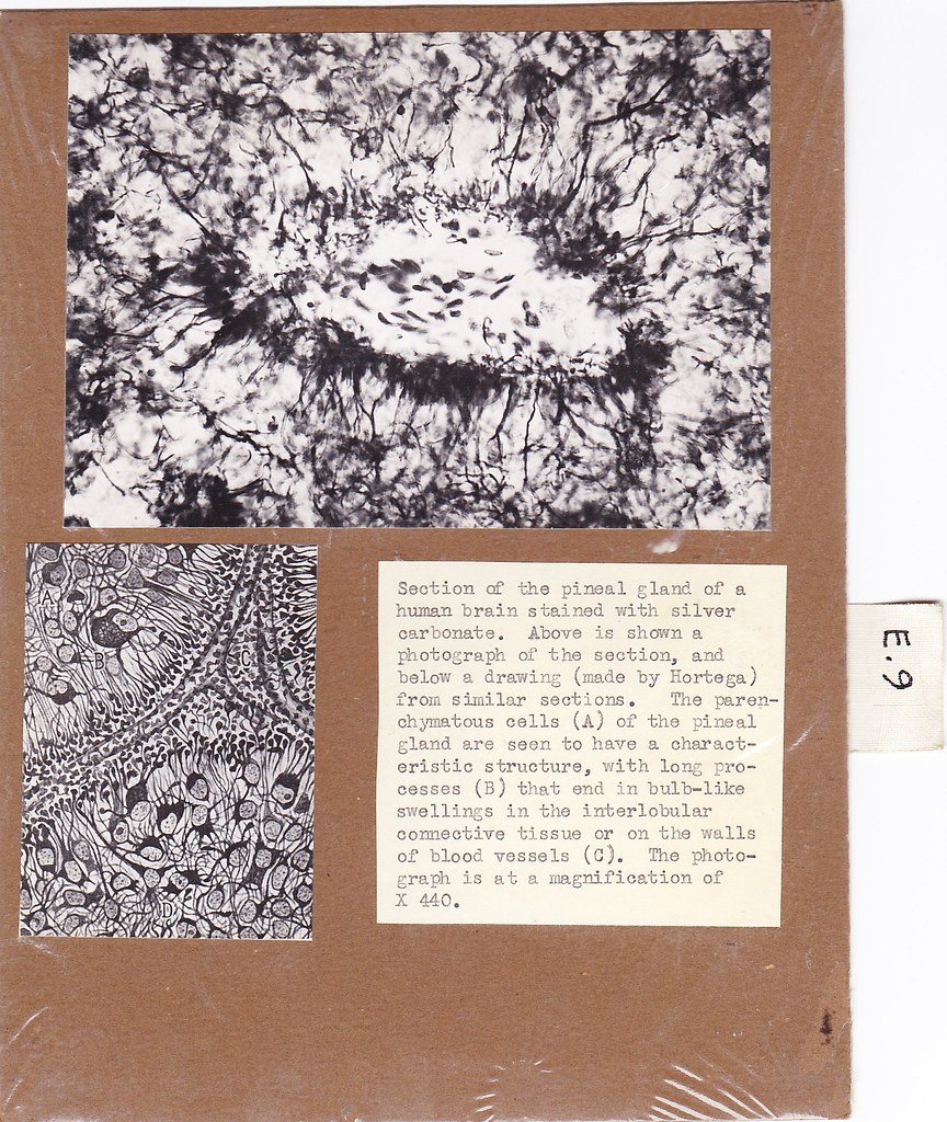
Section of the pineal gland of a human brain stained with silver carbonate. Above is shown a photograph of the section, and below a drawing (made by Hortega) from similar sections. The parenchymatous cells (A) of the pineal gland are seen to have a characteristic structure, with long processes (B) that end in bulb-like swellings in the interlobular connective tissue or on the walls of blood vessels (C). The photograph is at a magnification of X 440.
Original card


