- Tags
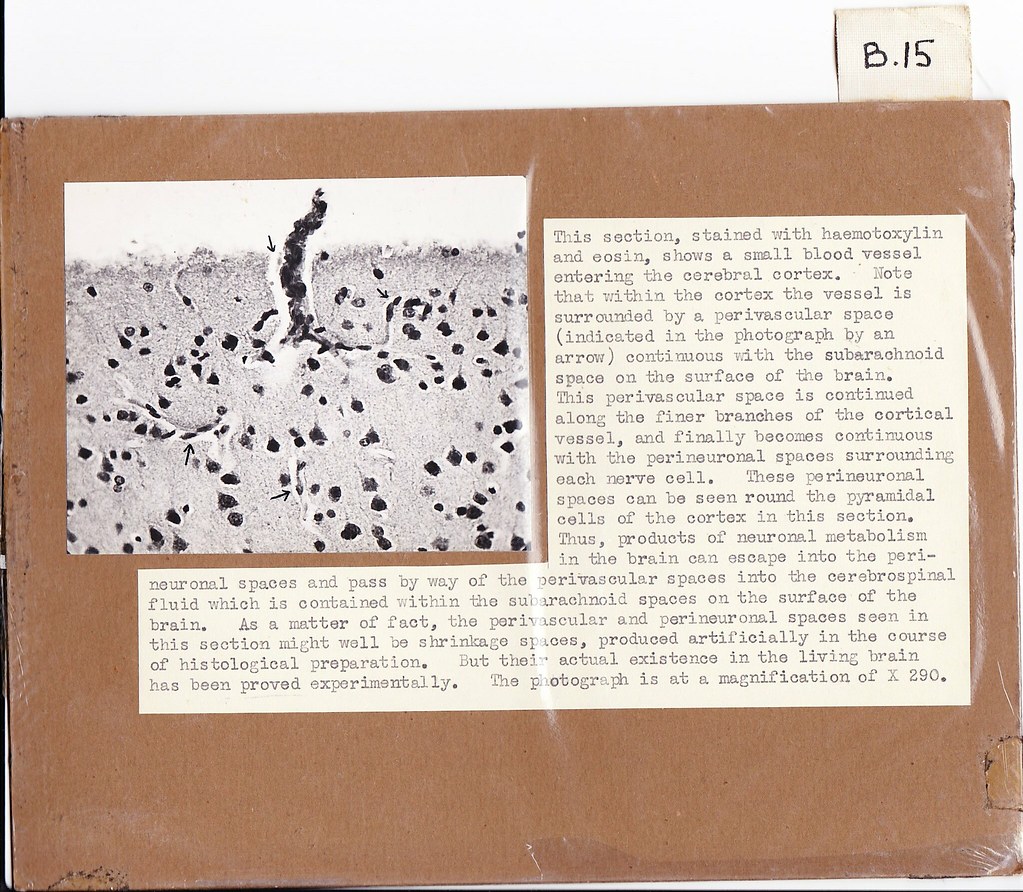
This section, stained with haemotoxylin and eosin, shows a small blood vessel entering the cerebral cortex. Note that within the cortex the vessel is surrounded by a perivascular space (indicated in the photograph by an arrow) continuous with the subarachnoid space on the surface of the brain. This perivascular space is continued along the finer branches of the cortical vessel, and finally becomes continuous with the perineuronal spaces surrounding each nerve cell. These perineuronal spaces can be seen round the pyramidal cells of the cortex in this section. Thus, products of neuronal metabolism in the brain can escape into the perineuronal spaces and pass by way of the perivascular spaces into fine cerebrospinal fluid which is contained within the subarachnoid spaces on the surface of the brain. As a matter of fact, the perivascular and perineuronal spaces seen in this section might well be shrinkage spaces, produced artificially in the course of histological preparation. But their actual existence in the living brain has been proved experimentally. The photograph is at a magnification of X 290.
Original card