- Tags
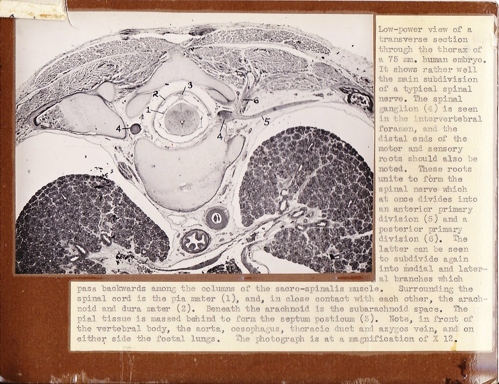
Low-power view of a transverse section through the thorax of a 75 mm. human embryo. It shows rather well the main subdivision of a typical spinal nerve. The spinal ganglion (4) is seen in the intervertebral foramen, and the distal ends of the motor and sensory roots should also he noted. These roots unite to form the spinal nerve which at once divides into an anterior primary division (5) and a posterior primary division (6). The latter can be seen to subdivide again into medial and lateral branches which pass backwards among the columns of the sacro-spinalis muscle. Surrounding the spinal cord is the pia mater (1), and, in close contact with each other, the arachnoid and dura mater (2). Beneath the arachnoid is the subarachnoid space. The pial tissue is massed behind to form the septum posticum (3). Note, in front of the vertebral body, the aorta, oesophagus, thoracic duct and azygos vein, and on either side the foetal lungs. The photograph is at a magnification of X 12.
Original card