- Tags
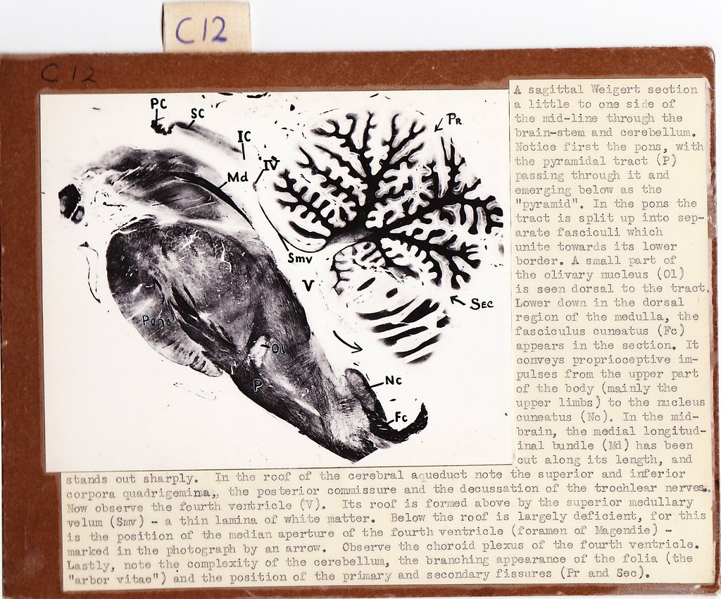
A sagittal Weigert section a little to one side of the mid-line through the brain-stem and cerebellum. Notice first the pons, with the pyramidal tract (P) passing through it and emerging below as the "pyramid". In the pons the tract is split up into separate fasciculi which unite towards its lower border. A small part of the olivary nucleus (Ol) is seen dorsal to the tract. Lower down in the dorsal region of the medulla, the fasciculus cuneatus (Fc) appears in the section. It conveys proprioceptive impulses from the upper part of the body (mainly the upper limbs) to the nucleus cuneatus (Nc). In the mid-brain, the medial longitudinal bundle (Md) has been cut along its length, and stands out sharply. In the roof of the cerebral aqueduct note the superior end inferior corpora quadrigemina, the posterior commissure and the decussation of the trochlear nerves. Now observe the fourth ventricle (V). Its roof is formed above by the superior medullary velum (Smv) − a thin lamina of white matter. Below the roof is largely deficient, for this is the position of the median aperture of the fourth ventricle (foremen of Magendie) − marked in the photograph by an arrow. Observe the choroid plexus of the fourth ventricle. Lastly, note the complexity of the cerebellum, the branching appearance of the folia (the "arbor vitae") and the position of the primary and secondary fissures (Pr and Sec).
Original card