- Creation
-
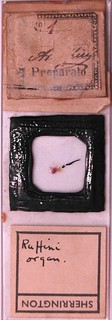
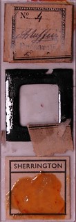
Creators (Definite): Dr Angelo Ruffini; Edwin Wilson
- Current Holder(s)
-

- No links match your filters. Clear Filters
-


-

-
 Cited byA. Ruffini, 'On the Minute Anatomy of the Neuromuscular Spindles of the Cat, and on their Physiological Significance', Journal of Physiology 23 (3) (1898), pp. 190-208.
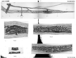
Description:Explanation of Plate III (figs. a-f and a-κ):
'Fig. a-f. Various forms of ordinary motor plates from the adult cat. These "plates" are figured from specimens taken from the same cat which furnished the preparations of the spindles represented in the preceding figures. Drawn as [Plate II] above.
Fig,. a-κ. Various types of "plate-endings" from those smaller (a, β, γ) to those larger than the common motor end-plates. These endings also have been drawn from preparations obtained from the same cat as that from which the motor end-plates a-f were taken. The preparations for both were obtained with the very same chloride of gold reaction.' (208)
Figs. a-f and a-κ in text:
'The plate-ending. Kerschner in 1888 noted this kind of ending and called it the motor apparatus, but nowhere has he ever given of it either description or figure. It was only after publication of my first Note that it became clear that Kerschner alluded to the kind of ending figured in Fig. 3 a of my Note. Besides an objective description of this ending I must give a comparison between it and ordinary motor endplates, not that from such a comparison a physiological function can be proved, but that from it a certain inference regarding function may be defensibly proposed.
The size of these plate-endings is most variable. Some are met with very much smaller than motor end-plates (Pl. III. a-γ), some about equal to them, and some very much larger than they; these last, the large ones, are the most common (cf. Pl. III.). From my own observations they appear to me unfurnished with granular supporting substance and with the Doyere eminence. The nerve-fibre which makes a plate-ending after it has laid aside its myelin sheath at the preterminal node breaks up into a few short, extremely delicate branched endings. These quickly begin to form what may be called coronets, by means of rounded varicose nerve-fibrils and very minute hooks set side by side. These coronets, which combine in various ways in the "plate," terminate by free ends which are obviously enlarged into bosses at the outer edge of the plate. By this arrangement arise a number of most delicate and elegant knobbed arborescences altogether different from the other two kinds of endings seen in the neuromuscular spindles. Rarely it can be seen that a pale fibre instead of an arborescence makes rather a web, in which however are constantly to be seen alternate widenings and narrowings of the naked nerve-fibre (PI. III. a-κ).' (202-203)
Figs. a-γ in text:
'Of primary endings usually only one can be found in each spindle. Rarely there are two, and then these lie widely apart. The Weissmann-bundle in the region of this ending is often slightly fusiform. Of the flower-spray enidings there are constantly a pair. These bear three different relations in respect to the primary ending: (a) one flowered ending lies on the proximal, another on the distal side of the primary ending, so that the primary is between them; (β) both flowered endings lie on the proximal side of the primary ending; (flowered endings lie on the proximal side of the primary ending; (γ) both flowered endings lie on the distal side of the primary ending. Whichever disposition occurs the closeness with which the primary and secondary endings keep together is curiously clear. The relation between them appears to me always contiguity not continuity.' (204-205)
Figs. a-f in text:
'The nerve-fibre after the preterminal node breaks up it is true into slender twigs, but these in forming the nervous-expansion never give the appearance of the coronet or the net but are short and thick, presenting here and there some irregular varicosities. The outer branches, often curved, give many or few side twigs that not unfrequently join one another, thus forming a network of extremely close mesh. The naked nerve-fibres of the motor plates also terminate with free but slightly widened ends (PI. III. a-f).' (203)
-
-
-
-
-
-


