- Creation
-
Creators (Definite): Walter Holbrook Gaskell; The Cambridge Scientific Instrument Company; Sir George Turner
- Current Holder(s)
-

- No links match your filters. Clear Filters
-


-

-

-
 Cited byW.H. Gaskell, 'On the Structure, Distribution and Function of the Nerves which innervate the Visceral and Vascular Systems', Journal of Physiology 7 (1) (1886), pp. 1-80.
Tags: osmic acid
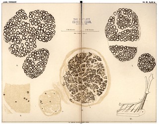
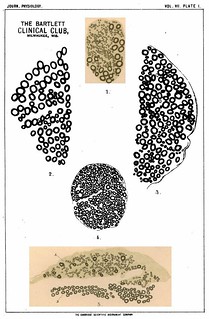
Description:Explanation of Plate IV (figs. 1-8):
'I am indebted to the kindness of Mr G. Turner for the figures of this plate. Figures 2, 3, 4, 5 were drawn from photographs taken by him of the microscopic sections.
Fig. 1 shows the connection between the posterior root ganglia and the sympathetic chain in the tortoise. Sy, sympathetic chain; the numbers 2-7 are placed against the corresponding thoracic nerves. The line l shows the direction of the series of parallel sections taken through the roots, ramus communicans, and root ganglion of the 5th thoracic nerve.
Figs. 2, 3, 4, 5 are copies of 4 sections out of this series and show the formation of ganglion cells on the sympathetic nerves, a, b, c, and the ultimate amalgamation of these ganglion cells with those of the root ganglion.
Fig. 6. Section of that branch of the 2nd thoracic nerve which forms the communicating branch to the 1st thoracic nerve and also helps to form the ramus visceralis of the ganglion stellatum. (From a photograph taken with lens 1/3 in.)
V. Small fibred portion of nerve which becomes the internal branch or ramus visceralis in connection with the ganglion stellatum.
A. Large fibred portion which becomes the external branch or branch of communication with the brachial plexus.
Fig. 7. Section of spinal accessory close to the ganglion jugulare vagi. (From a photograph taken with lens 1 in.)
V. Small fibred portion which becomes the internal branch or ramus visceralis in connection with the ganglion trunci vagi.
A. Large fibred portion which becomes the external branch and communicates with the cervical plexus.
Fig. 8. Upper part of spinal cord of dog cut in two in order to show the arrangement of the upper cervical nerve roots and those of the neural segment immediately above them.
1, 2, 3. Anterior and posterior roots of the corresponding cervical nerves.
V. Vagus nerve.
H. Hypoglossal nerve.
Ac. Spinal accessory nerve.' (79-80).
Fig. 1 in text:
'The connection of the fibres of the ramus visceralis with the cells of the ganglion on the posterior roots of the thoracic nerves is most clearly visible in the case of the tortoise. In this animal the ramus visceralis does not spring from the ventral branch of the spinal nerve as in mammalia, but arises directly from the ganglion on the posterior root. In Fig. 1, PI. IV., I give a representation of the connection between the sympathetic ganglia and the thoracic nerves from the 2nd to the 6th inclusive. As is seen, the ramus communicans directly connects each posterior root ganglion with its corresponding sympathetic ganglion. Owing to the inisertion of the lurmbo-caudalis muscle the rami communicantes of the 4th, 5th, 6th thoracic nerves at first pass out of the posterior root gangrlion in the direction of the roots of the nerves, and then turn over the muscle to reach the sympathetic chain.' (61-62)
Figs. 1-5 in text:
'In consequence of this arrangement, as is seen in Fig. 1, Pl. IV., a series of sections made for the purpose of following the spinal roots into the ganglion will at the same time enable us to trace the ramus communicans into the ganglion. In Figs. 2, 3, 4, 5, Pl. IV., I give drawings made from photographs of four sections taken from a consecutive series through the roots and spinal ganglion of the 4th thoracic nerve after staining with osmic acid. The figures show clearly how ganglion cells are formed in the ramus visceralis independently of those formed round the spinal roots, how these sympathetic ganglion cells increase in number as the posterior root ganglion is approached, and, finally, how they pass into and are lost in among the cells of the posterior root ganglion itself. Such a series makes it certain that in this case some of the nerve cells of the posterior root ganglion are connected with the fibres of the ramus visceralis.' (62)
Fig. 6 in text:
'The white ramus communicans of the 10th spinal nerve (2nd thoracic) is large and is composed of many separate bundles; the largest of these bundles arises directly-from this small branch of communication with the brachial plexus, and not from the main part of the 10th nerve. In Fig. 6, Pl. IV., I give a section of this communicating branch after it has left the main stem of the 10th (2nd thoracic) nerve. As is seen, it is composed of two parts, the one, A, chiefly containing large medullated fibres; the other, V, chiefly containing very fine medullated; as we trace the series of sections further and further from the origin of the nerve, we find the two portions separate more and more from each other'. (60)
Fig. 7 in text:
'In Pl. IV. Fig. 7, I give a picture of a section of the spinal accessory just before it reaches the ganglion jugulare of the vagus. The roots of the medullary portion of the accessory and of the vaaus were carefully hardened in situ with osmic acid, the whole with a portion of the medulla oblongata removed, imbedded in paraffin, and "ribbons" of consecutive sections made through the whole of the nerve roots from the medulla oblongata up to and beyond the ganglion trunci vagi. The whole series of sections was mounted in order, every nerve fibre was well stained and remained on the slide in the exact position it occupied when imbedded. In Pl. II. Fig. 10, the arrangement of the fibres when imbedded is reproduced.
The figure shows that the spinal accessory is divided into two distinct portions, the one (A) composed of large medullated fibres with a few isolated medium sized ones among them, the whole of that portion being remarkably free from connective tissue; the other portion (V) composed mainly of the smallest medullated fibres among which are a few large ones imbedded in a conspicuous matrix of connective tissue.' (10)
Fig. 8 in text:
'An examination of the relative positions of the roots of the nerve group immediately above the 1st cervical nerve (see Fig. 8, Pl. IV.) demonstrates the presence of two sets of rootlets which correspond in position to the anterior and posterior rootlets of the 1st cervical nerve, the only difference being that they are not so directly opposite to each other: a slight shifting of position which is easily accounted for by the alteration in the direction of the spinal axis due to the opening out of the central canal, and the formation of the 4th ventricle.' (74)
-
-