- Tags
- Creation
-
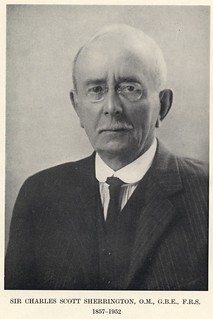
Creator (Possible): Sir Charles Scott Sherrington
- Current Holder(s)
-
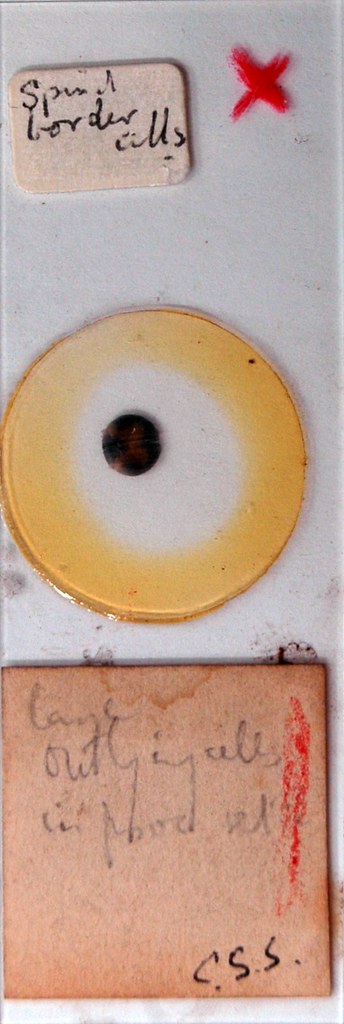
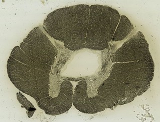
Spinal border cells from a Cooper and Sherrington publication from 1940.
Cooper, S. & Sherrington, C. S. On ‘Gower’s tract and spinal border cells’. Brain 68, 123–134 (1940).
Border cells were originally described by Gaskell23, who gave the name to the neurons scattered at the periphery of the lateral column in the spinal cord of the alligator.
In Sherrington’s box there are several spinal cord slides with labels pointing to cells at the edge of the spinal cord (or spinal border cells) (FIG. 1c,d in Molnar and Brown, 2010). These are the sections that led Sherrington24, while working at Cambridge and at St Thomas’ Hospital, to describe a group of large nerve cells in the ventrolateral grey matter of the lumbar spinal cord of monkeys and cats as ‘outlying nerve cells’. Later, in one of his last publications from the University of Oxford25, he called these neurons ‘spinal border cells’ because they were located predominantly along the lateral border of the ventral horn. Sherrington was interested in these cells because he suspected that they caused the sustained tonic inhibition of extensor muscle α-motor neurons in the cervical enlargement.
Only much later were these cells identified as spinocerebellar tract neurons26. Acute spinal injuries caudal to the cervical enlargement and cranial to border-cell neurons result in sudden deprivation of tonic inhibition of cervical enlargement neurons and cause their excitation. This excitation results in the extensor hypertonia observed in the thoracic limbs. Because Schiff27 described this syndrome in amphibian spinal cord before Sherrington, it is usually referred to as the Schiff–Sherrington phenomenon28. This work provides an excellent example of the way Sherrington combined anatomical and physiological approaches to understand the interactions among spinal circuits that regulate reflex action by inhibition.
- No links match your filters. Clear Filters


