- Creation
-
Creators (Definite): Sir John Scott Burdon-Sanderson; Prof Francis GotchDate: 1888
- Current Holder(s)
-
From article:
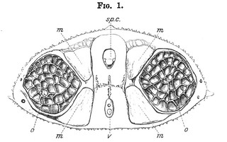
Caption: 'Longitudinal section of upper part of organ as seen under low power (Raia batis). The arrow indicates the direction of the shock.' (142)
'In fig. 2 the engraver has given the general effect of a photograph of a longitudinal frontal section, as seen under the microscope with a low power. It serves to shew that the organ consists of spindle-shaped tubes imperfectly divided into loculi placed one above the other, and each holding a disk. These tubes are so arranged that their axes are either parallel or very slightly diverge backwards. The disks are, so to speak, suspended by the connective tissue which supports the blood-vessels. As has already been stated, the arterioles follow in the first instance the longitudinal septa by which the tubes are separated from each other. From the septa terminal arterioles pass transversely, i.e. at right angles to the axis of each tube, into the spaces between each two adjoining disks, occupying a position about half way between their two opposed surfaces. The description of their distribution, which we have given from M. Robin's Memoir, need not be repeated. Both arteries and veins are accompanied by connective tissue, which in the horizontal part of their course is of sufficient strength to deserve the name of a lamina, although it is distinguished from the rest of the connective tissue which occupies the space between the disks merely by the closer arrangement of its fibres.' (141-142)

- No links match your filters. Clear Filters
-


-
-
-